Download
1 / 8
80 likes | 251 Views
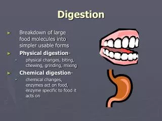
Digestion. Small Intestine & Pancreas. Structure of the Small Intestine. The S.I. is about 2.5 cm in diameter and 7 m in length and made up of 3 segments: Duodenum Jejunum Ileum The S.I. is made of the same 4 layers found in the stomach: Serosa, muscle, submucosa , and mucosa

E N D
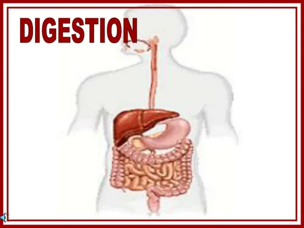
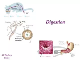
Digestion Small Intestine & Pancreas
Structure of the Small Intestine • The S.I. is about 2.5 cm in diameter and 7 m in length and made up of 3 segments: • Duodenum • Jejunum • Ileum • The S.I. is made of the same 4 layers found in the stomach: Serosa, muscle, submucosa, and mucosa • The main function of the S.I. is digestion and absorption of nutrients
Structure (cont) • Inner surface of S.I. is adapted to provide maximum surface area for nutrient absorption. • Inner surface is folded into ridges (rugae) • Rugae coated in finger-like projections (villi) • The cells lining each villus have even smaller projections (microvilli)
Structure (cont) • Each villus contains blood vessels and a lacteal • All nutrients, except fats, enter the bloodstream through the capillaries • Digested fats enter through lacteals into the lymphatic system before going to the blood stream
Chemical Digestion in The S. I. • Most of the enzymes for digestion are added in the duodenum. • Many of these enzymes are supplied by the liver, gall bladder, and pancreas. Fig. 3 Secretions enter intestine through ducts that connect each organ to the duodenum
Role of the Pancreas • Cholecystokinin (CCK) – hormone released by intestines when chyme, that is high in fat, enters the blood stream • Signals stomach to slow emptying • Signals pancreas to release enzymes • Pancreatic Amylase – continues starch digestion started by saliva • Insulin/Glucagon – hormones that regulate blood sugar • Insulin helps cells remove glucose from blood • Glucagon releases glucose to the blood from the liver
Role of the Pancreas (cont) • Prosecretin – hormone produced in the S.I. • activated to ‘secretin’ when it contacts acidic chyme from stomach. • Secretin signals pancreas to release bicarbonate ions (HCO3-) to neutralize acidic chyme • pH 2.5 9.0 and deactivates pepsin • Secretin also signals pancreas to release trypsinogenand erepsinto breakdown protein and lipases to break down fat
Protein Digestion in S.I Enterokinase (from S.I.) Trypsinogen (from pancreas) Trypsin Erepsin (from pancreas) Amino Acids Long-Chain Proteins Shorter-Chain Proteins