Download
1 / 47
470 likes | 499 Views
Learn the diagnostic process for Undifferentiated Peripheral Inflammatory Arthritis through a case study. Explore history-taking, physical examination, laboratory tests, and antibody evaluations for accurate diagnosis and prognosis assessment. Receive expert recommendations and insights for managing UPIA effectively.

E N D
3e Initiative 2009How to investigate and follow-up Undifferentiated Peripheral Inflammatory Arthritis? Case 1
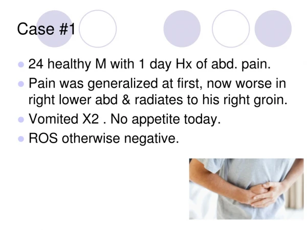
Case 1 • Elisa, an art teacher and active smoker comes to your consultation because of a swelling of the left knee and the right 4th proximal (ring finger) interphalangeal joint • Symptoms began 2 months ago with tenderness of both hands • She has no family history • Her Hashimoto’s disease is treated by L-thyroxine and at present is well controlled
Question 1 • In your opinion, which features on history may help to predict diagnosis of rheumatoid arthritis? • Her age • Her reproductive history • A recent infection history • The duration of morning stiffness • Self report of pain
Answer Question 1 • In your opinion, which features on history may help to predict diagnosis of rheumatoid arthritis? • Her age • Her reproductive history • A recent infection history • The duration of morning stiffness • Self report of pain
Question 2 • In your opinion, which features on physical exam may help to predict a diagnosis of rheumatoid arthritis? • Tender Joint Count • Swollen Joint Count • Joint distribution • Stress Pain • Joint symmetry • Body Mass Index
Answer Question 2 • In your opinion, which features on physical exam may help to predict diagnosis of rheumatoid arthritis? • Tender Joint Count • Swollen Joint Count • Joint distribution • Stress Pain • Joint symmetry • Body Mass Index
Recommendation 2 • To establish a specific diagnosis and prognosis following presentation of UPIA, a careful systematic history and physical examination should be performed, with particular attention to: • Age • Gender [1a, A] • Geographic area [5, D] • Functional status [1a, A] • Duration of symptoms / early morning stiffness • Number plus pattern of tender / swollen joints [1a, A] • Axial / entheseal involvement • Extra-articular / systemic features [5, D]
Historical features with diagnostic utility for the development of RA *Estimates did not cross null value of no association *
Physical exam features found to have diagnostic utility for the development of RA *Estimates did not cross null value of no association *
Question 3 • She is 51 years old and has the following disease activity parameters: • Morning stiffness >1 hour • Tender joints: 4 • Swollen joints: 2 • You are planning to perform laboratory tests to help classify this arthritis • In your opinion, which acute phase reactants have some diagnostic value for defining RA? • ESR • CRP • Ferritin • Matrix MetalloProteinase 3 (MMP) • Complete Blood Count
Answer Question 3 • She is 51 years old and has the following disease activity parameters: • Morning stiffness >1 hour • Tender joints: 4 • Swollen joints: 2 • You are planning to perform laboratory tests to help classify this arthritis • In your opinion, which acute phase reactants have some diagnostic value for defining RA? • ESR • CRP • Ferritin • Matrix MetalloProteinase 3 (MMP) • Complete Blood Count
Recommendation 3 • ESR and CRP should be performed at baseline in the work up for diagnosis [2b, B], and prognosis [2b, B], of UPIA and repeated when clinically relevant [5, D]
Question 4 • Which antibodies will help predict the development of RA? • Rheumatoid factor • ANA • ACPA (anti-CCP) • ANCA • Anti-MCV • Anti-RA33
Answer Question 4 • Which antibodies will help predict the development of RA? • Rheumatoid factor • ANA • ACPA (anti-CCP) • ANCA • Anti-MCV • Anti-RA33
Recommendation 4 • Testing of RF and/or ACPA should be performed in the evaluation of patients with UIPA, as these factors are predictive of RA diagnosis and prognosis; negative tests do not exclude progression to RA [1a, A] • If a connective tissue disease / systemic inflammatory disorder is suspected, additional autoantibody tests should be considered [5, D]
Antibodies: diagnostic value for prediction ofRA at 1 to 3 years • RF is consistently associated with the diagnosis of RA (LR+ 1.1 to 13.5) • The absence of RF is diagnostically less helpful (LR- 0.3 to 0.8) • Anti-CCP is consistently associated with the diagnosis of RA (LR+ 1.2 to 20.5) • The absence of anti-CCP is diagnostically less helpful (LR- 0.4 to 0.9)
Question 5 • You want to order radiographs. Which one(s) should you request? • Right hand radiograph • Both hands and wrists radiographs • Feet radiographs • Left knee radiograph • Pelvic/sacroiliac joints radiographs
Answer Question 5 • You want to order radiographs. Which one(s) should you request? • Right hand radiograph • Both hands and wrists radiographs • Feet radiographs • Left knee radiograph • Pelvic/sacroiliac joints radiographs
Question 6 • She comes back with her X-ray. Which pathological features will help you to predict the development of RA? • Bony decalcification • Radiographic erosion • Joint space narrowing • Calcifications • Enthesophytes
Answer Question 6 • She comes back with her X-ray. Which pathological features will help you to predict the development of RA? • Bony decalcification • Radiographic erosion • Joint space narrowing • Calcifications • Enthesophytes
Recommendation 5 (beginning) • Radiographs of affected joints should be performed at baseline [5, D] • Radiographs of hands, wrists, and feet should be considered in the evaluation of UPIA, as presence of erosions is predictive for the development of RA and persistence of disease [1a, A] • […]
Question 7 • The results of the lab tests and radiographs are the following: • ESR: 25 mm at 1st hour • CRP: 30 mg/L • RF negative • Anti-CCP negative • Normal X-ray • How do you interpret these results so far in making a diagnosis? • Reconsider diagnosis of RA • Reconsider diagnosis of UPIA • Match progression to RA • Match diagnosis of UPIA
Answer Question 7 • The results of the lab tests and radiographs are the following: • ESR: 25 mm at 1st hour • CRP: 30 mg/L • RF negative • Anti-CCP negative • Normal X-ray • Do these results ESR • Reconsider diagnosis of RA • Reconsider diagnosis of UPIA • Match progression to RA • Match diagnosis of UPIA
Question 8 • The results of the lab tests and radiographs are the following: • ESR: 25 mm at 1st hour • CRP: 30 mg/L • RF negative • Anti-CCP negative • Normal X-ray • How often will you repeat these exams? • Never • Acute phase reactants once a month • Auto-antibodies every three months • According to the clinical setting • Radiographs not before 2 years
Answer Question 8 • The results of the lab tests and radiographs are the following: • ESR: 25 mm at 1st hour • CRP: 30 mg/L • RF negative • Anti-CCP negative • Normal X-ray • How often will you repeat these exams? • Never • Acute phase reactants once a month • Auto-antibodies every three months • According to the clinical setting • Radiographs not before 2 years
Recommendations 3, 4 and 5 • ESR and CRP should be [...] repeated when clinically relevant [5, D]No data available concerning the fact whether acute phase reactants should be repeated at certain time-intervals • […] RF and/or anti-citrullinated negative tests ACPA do not exclude progression to RA [1a, A]The current evidence does not allow to make conclusions on the optimal frequency of serological assessments in the evaluation of patients with UPIA. • Radiographs [...] should be repeated within one year [5, D]No data was found about the value of repeating X-rays in UA patients or mixed populations
Question 9 • Elisa comes back to your consultation 2 months later. In spite of analgesic and NSAIDs you gave her, she has a a pain and stiffness of her right wrist and a persistent swelling of the right 4th proximal interphalangeal joint. Considering results of her first exams, which imaging investigations can you propose to her to confirm a potential diagnosis of RA? • New hands and wrists radiographs • Right hand and wrist Magnetic resonance imaging (MRI) • Hands and Wrists ultra-sound (US)
Answer Question 9 • Elisa comes back to your consultation 2 months later. In spite of analgesic and NSAIDs you gave her, she has a a pain and stiffness of her right wrist and a persistent swelling of the right 4th proximal interphalangeal joint. Considering results of her first exams, which imaging investigations can you propose to her to confirm a potential diagnosis of RA? • New hands and wrists radiographs • Right hand and wrist Magnetic resonance imaging (MRI) • Hands and Wrists ultra-sound (US)
Recommendation 6 and 5 • There is insufficient evidence to recommend the routine use of magnetic resonance imaging (MRI) and ultra-sound (US) for diagnosis or prognosis in UPIA [5, D]; in UPIA and suspicion of RA, MRI of hands and wrists could be considered for diagnosis [2b, B] • […] Radiographs should be repeated within one year [5, D]
Diagnostic value of US: mixed population NB: In patients seronegative for RF and CCP BUT positive for CRP, swollen joints and X-ray erosion, the presence of US GS=3 , US PD≥1 and at least 1 erosion increased the probability of IA from 30% to 94%. If only 1 US feature was positive, the probability of IA was 0-39% If 2 US features were positive it was 8-85% If all US features were negative, the probability of IA was only 0-5%.
Question 10 • MRI shows bone edema on her right wrist. She asks you how long she will suffer from this arthritis. Which elements in this observation are predictors of persistent inflammatory arthritis? • Disease duration (4 months) • Morning stiffness (>1 hour) • Swelling of 2 joints • Involvement of the knee • ESR: 25 mm at 1st hour and CRP: 30 mg/L • Bone edema on IRM
Answer Question 10 • MRI shows bone edema on her right wrist. She asks you how long she will suffer from this arthritis. Which elements in this observation are predictors of persistent inflammatory arthritis? • Disease duration (4 months) • Morning stiffness (>1 hour) • Swelling of 2 joints • Involvement of the knee • ESR: 25 mm at 1st hour and CRP: 30 mg/L • Bone edema on IRM
Recommendation 9 Predictors of persistent inflammatory arthritis should be documented and include disease duration of ≥6 weeks [1b, A], morning stiffness >30 min [4, C], functional impairment [4, C], involvement of small joints [4, C] and/or knee [4, C], ≥ 3 joints [1b, B], ACPA [4, C] and/or RF positivity [4, C] and presence of radiographic erosion [1b, B].
Predictors of persistent (chronic) UPIA: Study Characteristics * Synovitis = 2 of following 3 criteria :- swelling, tenderness, and decreased range of motion
Predictors of persistent disease: Summary Oxford level of evidence :- 1b
Question 11 • You would like to evaluate the disease activity to better manage the follow-up. Which are the most suitable clinical measures (any activity index, measure or scales) to evaluate the diagnosis and the follow-up of UPIA? • DAS (Disease Activity Score) • CDAI (Clinical Disease Activity Index) • SDAI (Simplified Disease Activity Index) • WHODAS II (WHO Disability Assessment Schedule) • HAQ (Health Assessment Questionnaire) • McROMI (McGill Range of Motion Index) • RADAI (RA Disease Activity Index) • No specific tool can be recommended
Answer Question 11 • You would like to evaluate the disease activity to better manage the follow-up. Which are the most suitable clinical measures (any activity index, measure or scales) to evaluate the diagnosis and the follow-up of UPIA? • DAS (Disease Activity Score) • CDAI (Clinical Disease Activity Index) • SDAI (Simplified Disease Activity Index) • WHODAS II (WHO Disability Assessment Schedule) • HAQ (Health Assessment Questionnaire) • McROMI (McGill Range of Motion Index) • RADAI (RA Disease Activity Index) • No specific tool can be recommended
Recommendation 10 • Disease activity should be monitored [no evidence, D], however no specific tool can be recommended [3b, C] • No instrument of disease activity has been fully validated for its use in UPIA • Specifically the more frequent indexes, such as DAS28, SDAI, SDAI... • Literature search has not found any direct evidence on neither what is the most useful index to follow up patients with UPIA, nor, obviously, at which interval we should repeat it
Activity measures in UPIA: Summary of the results Questionnaires Indexes