ELECTROENCEPHALOGRAPHY (EEG)
ELECTROENCEPHALOGRAPHY (EEG). Dr. Shaikh Mujeeb Ahmed M.B.B.S. MD. (PHYSIOLOGY). EEG. The electroencephalogram (EEG) is a recording of the electrical activity of the brain from the scalp. The first recordings were made by Hans Berger in 1929 . Origin of EEG waves. Electroencephalogram.


ELECTROENCEPHALOGRAPHY (EEG)
E N D
Presentation Transcript
ELECTROENCEPHALOGRAPHY(EEG) Dr. Shaikh Mujeeb Ahmed M.B.B.S. MD. (PHYSIOLOGY)
EEG • The electroencephalogram (EEG) is a recording of the electrical activity of the brain from the scalp. • The first recordings were made by Hans Berger in 1929
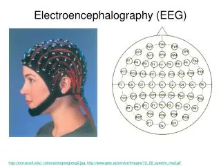
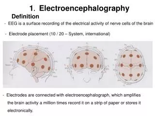
Electroencephalogram • EEG is the record of electrical activity of brain( superficial layer i.e. the dendrites of pyramidal cells) by placing the electrodes on the scalp.
Objectives of EEG practical • Familiarize with the principles of techniques involved • Count frequencies and measure the amplitudes of the record obtained. • Categories the records into appropriate rhythms – α, β, θ,and δ. Cont…
Objectives of EEG practical • Identify and describe changes produced by provocation tests. e.g. eye opening & closing, intermittent photic stimulation (IPS) clapping sound, induce thinking & hyperventilation. • Appreciate clinical uses of EEG
EEG Waves • Alpha wave -- 8 – 13 Hz. • Beta wave -- >13 Hz. (14 – 30 Hz.) • Theta wave -- 4 – 7.5 Hz. • Delta waves – 1 – 3.5 Hz. • D T A B
Alpha wave • rhythmic, 8-13 Hz • mostly on occipital lobe • 20-200 μ V • normal, • relaxed awake rhythm with eyes closed
Beta wave • irregular, 14-30 Hz • mostly on temporal and frontal lobe • mental activity • excitement
Theta wave • rhythmic, 4-7 Hz • Drowsy, sleep
Delta wave • slow, < 3.5 Hz • in adults • normal sleep rhythm
Requirements • EEG machine (8/16 channels). • Silver cup electrodes/metallic bridge electrodes. • Electrode jelly. • Rubber cap. • Quiet dark comfortable room. • Skin pencil & measuring tape.
EEG Electrodes Electrodes Cap Sliver Electrodes
Procedure of EEG recording • A standard EEG makes use of 21 electrodes linked in various ways (Montage). • Ask the subject to lie down in bed. • Apply electrode according to 10/20% system. • Check the impedance of the electrodes.
Procedure of EEG recording • Ask the subject to close his/her eyes. • Select a montage. • Press run switches on to run the paper.
Procedure of EEG recording • Press the calibration knob to check voltages & time constant. • Always observe subject for any abnormal muscle activity. • Ask the subject to open eyes for 10 sec.and ask him/her to close eyes. (do this procedure for several times in each montage)
EEG Electrodes • Each electrode site is labeled with a letter and a number. • The letter refers to the area of brain underlying the electrode e.g. F - Frontal lobe and T - Temporal lobe. • Even numbers denote the right side of the head and • Odd numbers the left side of the head.
Two types of recording • Bipolar – both the electrodes are at active site • Bipolar montage are parasagital montage. • Unipolar – one electrode is active and the other is indifferent kept at ear lobe. • Always watch for any abnormal muscle activity. • Ask the subject to open eyes for 10 sec. then ask them to close the eyes.
Montage • Different sets of electrode arrangement on the scalp by 10 – 20 system is known as montage. • 21 electrodes are attached to give 8 or 16 channels recording.
Analysis • Electrical activity from the brain consist of primarily of rhythms. • They are named according to their frequencies (Hz) and amplitude in micro volt (μv). • Different rhythms at different ages and different conditions (level of consciousness) • Usually one dominant frequency (background rhythm)
Factor influencing EEG • Age • Infancy – theta, delta wave • Child – alpha formation. • Adult – all four waves. • Level of consciousness (sleep) • Hypocapnia(hyperventilation) slow & high amplitude waves. • Hypoglycemia • Hypothermia • Low glucocorticoids Slow waves
Desynchronization or Alpha block • Cause: • Eyes opening (after closure) • Thinking by the subject (mathematical calculation) • Sound (clapping)
Eye opening • Alpha rhythm changes to beta on eye opening (desynchronization / α- block)
Thinking • Beta waves are observed
Provocation test • Intermittent photic stimulation • Increase rate & decrease amplitude • Hyperventilation • Decrease rate & increase in amplitude
Use of EEG • Epilepsy • Generalized (grandmal) seizures. • Absence (petitmal) seizures. • Localize brain tumors. • Sleep disorders (Polysomnography) • Narcolepsy • Sleep apnea syndrome • Insomnia and parasomnia • Helpful in knowing the cortical activity, toxicity, hypoxia and encephalopathy & • Determination of brain death. • Flat EEG(absence of electrical activity) on two records run 24 hrs apart.
Sleep studies • The EEG is frequently used in the investigation of sleep disorders especially sleep apnoea. • Polysomnography : EEG activity together with • heart rate, • airflow, • respiration, • oxygen saturation and • limb movement
Sleep patterns of EEG • There are two different kinds of sleep: • Rapid eye movement sleep (REM-Sleep) • Non-REM sleep (NREM sleep)/ slow wave sleep • NREM sleep is again divided into 4 stages (I to IV). The EEG pattern in sleep is given in the following table:
Changes in brain waves during different stages of sleep & wakefulness
Changes in brain waves during different stages of sleep & wakefulness
K - complex Sleep Spindle