Electroencephalography & Event-related potentials ( EEG & ERP)
810 likes | 1.23k Views
Electroencephalography & Event-related potentials ( EEG & ERP). علی یونسی پژوهشکده علوم شناختی اردیبهشت 91. مصرف کننده شیشه (متآمفتامین). مصرف کننده قبلی شیشه. فرد سالم. خلاصه. مقدمه ای بر نوار مغزی و سیگنال ناشی از محرک منشا فعالیت الکتریکی مغز ریتمهای مغزی حالات مغز کاربردهای EEG

Electroencephalography & Event-related potentials ( EEG & ERP)
E N D
Presentation Transcript
Electroencephalography & Event-related potentials (EEG & ERP) علی یونسی پژوهشکده علوم شناختی اردیبهشت 91
مصرف کننده شیشه (متآمفتامین) مصرف کننده قبلی شیشه فرد سالم
خلاصه • مقدمه ای بر نوار مغزی و سیگنال ناشی از محرک • منشا فعالیت الکتریکی مغز • ریتمهای مغزی • حالات مغز • کاربردهای EEG • کاربردهای شناختی • یادگیری • حافظه • توجه • تفسیر رویا • کاربردهای بالینی • تشنج • اختلالات حافظه • پایش • پایش تشنج • پایش در طول بیهوشی • پایش در حین عمل اندارترکتومی
کاربردهای شناختی ERP • BCI • ورودیهای حسی • خروجیهای حرکتی • تفسیرهای عملکردی • توجه • یادگیری • کاربردهای بالینی ERP • Schizophrenia • Mood disorders • Alcohol dependence and substance abuse • Dementia • Traumatic brain injury • Normal development • Childhood disorders • ADHD • Learning Disorders • تحقیقات در حال انجام • سوء مصرف شیشه • خواب
Research and Application • Psychological Research • Neurological Research • Medical Research • Educational Research and Application • Therapeutic Application • Occupational Application
پوست بافت نرم جمجمه دورا کورتکس 5 mm 5 mm ثبت سیگنال • الکتریکی • Electroencephalography (EEG) • Electrocorticogram (ECoG) • Local field potential or single neuron • روشهای دیگر • Magnetoencephalography (MEG) • Positron emission tomography (PET) • Magnetic resonance imaging (fMRI) • Infrared (IR) imaging
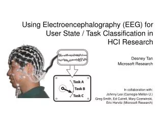
How do EEGs work? Neural communication produces electrical activity. Electrical activity in a single neuron. When a neuron is active, its voltage may change by 100 mV or more.
Basics: Inhibition, Amplitude and Timing Large amplitudes tend to entrain many neurons Cell 1 Cell 2 Excitation (Pyramidal cells) Maximum Minimum Inhibition Minimum Maximum Cell 3 Cell 1 Cell 2 Cell 3 Time
Cortical Basis of Scalp EEG Baillet et al., IEEE Sig. Proc. Mag., Nov 2001, p. 16.
Mountcastle, Brain, 120:701-722, 1997. Six Layer Cortex
EEG Electrodes Electrodes Cap Sliver Electrodes
How do EEGs work? This activity may be detectable to electrodes on the scalp. Conventional electrode caps from EGI, Neuroscan, and Electro-Cap.
برتریهای نسبی به دیگر روشها • Hardware costs are significantly lower than those of all other techniques • EEG sensors can be used in more places than fMRI, SPECT, PET, MRS, or MEG, • EEG has higher temporal resolution - milliseconds, rather than seconds - it can, in fact, take as many as 2000 samples per second (Only MEG rivals these speeds) • EEG is relatively tolerant of subject movement • EEG is silent • EEG does not aggravate claustrophobia • EEG does not involve exposure to high-intensity (>1 Tesla) magnetic fields • ERP studies can be conducted with relatively simple paradigms, compared with block-design of fMRI studies • Extremely uninvasive
نقصان نسبت به دیگر روشها • Significantly lower spatial resolution. fMRI, for example, can directly display areas of the brain that are active, while EEG requires intense interpretation just to hypothesize what areas are activated by a particular response. • EEG determines neural activity that occurs below the upper layers of the brain (the cortex) very poorly. Unlike PET and MRS, cannot identify specific locations in the brain at which various neurotransmitters, drugs, etc. can be found. • Often takes a long time to connect a subject to EEG, as it requires precise placement of dozens of electrodes around the head and the use of various gels, saline solutions, and/or pastes to keep them in place. • Signal-to-noise ratio is very poor, so sophisticated data analysis and relatively large numbers of subjects are needed to extract useful information from EEG
چند نکته حیاتی • a given electrode on the scalp does not record solely the neuronal activity directly underlying it. Rather, every electrode picks up signals from different sources that can eventually be quite distal • Fluctuation of the voltage at the reference electrode will lead to changes of the potential at the active electrode even if the voltage at that point was actually stable. There is no point that is electrically silent and could be considered as true zero potential.
Midline Fronto-central Centro-parietal Anterior Posterior
Alpha wave • rhythmic, 8-13 Hz • mostly on occipital lobe • 20-200 μ V • normal, • relaxed awake rhythm with eyes closed
Beta wave • irregular, 14-30 Hz • mostly on temporal and frontal lobe • mental activity • excitement
Theta wave • rhythmic, 4-7 Hz • Drowsy, sleep
Delta wave • slow, < 3.5 Hz • in adults • normal sleep rhythm
EEG recording in man • Eyes opened condition. • Examples of different waves.
The cocktail party problem - find Z x1 z1 x2 z2 A xN XT=AZT zN ZT XT
Blind Source Separation Blind Source Separation deals with the « separation » of a mixture of sources, witha littleprior information about the mixingprocess and the sources signals x = As Ŝ = Wx Environment x1 Ŝ 1 S1 S2 SN Source Separation Algorithm W x2 Ŝ2 . . . . . . . . . . . . xN ŜN Sources Sensors Observations
Nyquist Theorem • The highest frequency which can be accurately represented is one-half of the sampling rate. • The sampling rate here is below the Nyquist frequency, so the result of sampling is nothing like the input:aliasing. • For practical purposes the sampling rate should be 10 higher than the highest frequency in the signal.
LORETA One such method is known as "LORETA", which provides an estimate of the current distribution throughout the entire 3-dimensional space within the brain. An example of a LORETA solution, mapped onto a normalized brain space, is provided below. SPAN 4130 - Harry Howard - Tulane University
Brain Wave Activity • Delta – sleep state (1-3 Hz) • Theta – between sleep and awake (4-7 Hz) • Alpha – relaxed state (8-12 Hz) • Low Beta – focused concentration (SMR-Sensory Motor Rhythms) (12-15 Hz) • Mid-range Beta – alert state (15-18 Hz) • High Beta – very alert, vigilant (Above 18) • Gamma – Hyper vigilant (Above 40)
Default-mode brain • The default network is a network of brain regions that are active when the individual is not focused on the outside world and the brain is at wakeful rest. • Working memory tasks differentially deactivate the PCC. • signal increase and spatial decrease in the PCC and a signal decrease but spatial increase in the ACC with increasing working memory load
Oscillations and the control of information processing (1) Oscillations: Timing and spatial organization of information processes. Oscillations provide mechanisms that allow the emergence of spatially and temporally organized firing patterns in neural networks. (2) Slow frequency oscillations: Conscious control of information processing.Slow frequency oscillations in the theta and alpha range (of about 4 – 13.5 Hz) are associated with the top-down control of two large processing systems, a working memory system and a a complex knowledge system, allowing semantic orientation in a constantly changing environment. Theta and alpha oscillations exhibit a variety of different synchronization processes (e.g., amplitude increase, phase coupling, event-related phase reorganization) that reflect different types of control processes and different aspects of the timing of cognitive processes.
Example of an evoked, ‘traveling’ theta wave, one subject, negative polarity is in blue A Fz Theta-waves single subject “B“ Cz -1.5 po p -1 p+1 Pz -1.0 Oz -0.5 µV 0.0 0.5 1.0 change in direction 1.5 500 600 700 800 900 1000 1100 1200 1300 ms poststim
برانگیختگی ناشی از محرک • کاربردها • ترجمه سیگنال به یک حرکت • استفاده از مغز به عنوان یک پردازشگر سریع • بررسی عملکرد حسی و شناختی فرد
Alzheimer’s disease and mild cognitive impairment • Semantic classification task: low frequency functional connectivity between anterior (MPFC) and posterior (PCC/retrosplenial cortex) regions : negatively associated with age • eyes-closed resting state in Alzheimer patients (N=24; 9 males; mean age 76.3 years)and non-demented subjects with subjective memory complaints (N=19; 9 males) • The mean level of EEG synchronization was lower in Alzheimer patients in the upper alpha (10–13 Hz) and beta (13–30 Hz) band.
Schizophrenia • low-frequency and alpha-band power abnormalities (perhaps thalamic and frontal lobe dysfunction) • augmented low-frequency power : • more negative symptoms • larger third ventricles • larger frontal horns of the lateral ventricles • increased cortical sulci widths • greater ocular motor dysfunction
Autism • In the θ (3–6 Hz) frequency range • within left hemisphere frontal and temporal regions • 8–10 Hz: • globally reduced coherence within frontal regions and between frontal and all other scalp regions. • The ASD : greater relative power between 3 and 6 Hz and 13–17 Hz and significantly less relative power between 9 and 10 Hz.
برانگیختگی بدون محرک هدف برانگیختگی با محرک هدف خصوصیات نورونی • برانگیختگی ناشی از محرک • P3 یکی از بخشهای خاص این سیگنال برانگیخته است: • میزان توجه • سختی پردازش تصویر • سن فرد