Download
1 / 11
130 likes | 317 Views
Explore the histology, function, and digestion processes of the small intestine and liver, including the role of bile in digestion and metabolism. Learn about the different regions and functions of the small intestine.

E N D
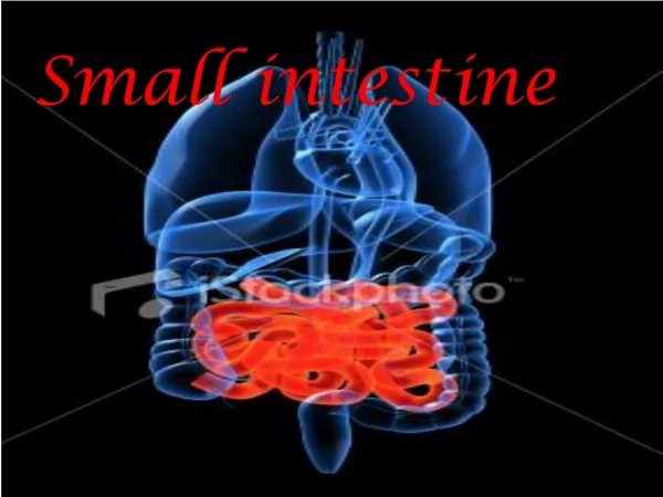
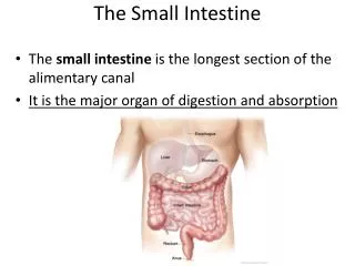
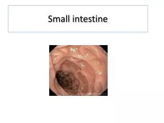
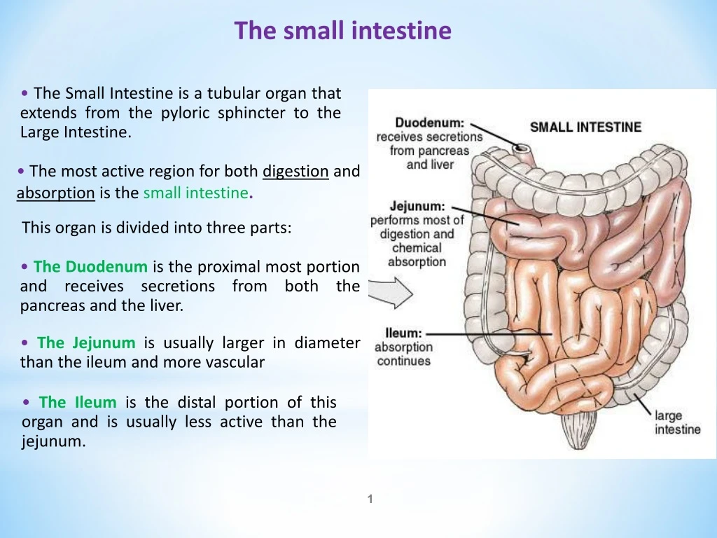
The small intestine • The Small Intestine is a tubular organ that extends from the pyloric sphincter to the Large Intestine. • The most active region for both digestion and absorption is the small intestine. This organ is divided into three parts: • The Duodenum is the proximal most portion and receives secretions from both the pancreas and the liver. • The Jejunum is usually larger in diameter than the ileum and more vascular • • The Ileum is the distal portion of this organ and is usually less active than the jejunum.
The histology of the small intestine •The interior lining of the intestinal wall are covered by numerous Villi. •These structures project into the lumen of the intestine and greatly increase the absorptive capacity of this organ. •Eachvillusis made up of a layer of epithelium and a core of connective tissue containing blood capillaries, nerve fibers, and a lymphatic Lacteal. •At their free surface the epithelial cells have many fine extensions called Microvillithat form what is known as the “Brush Boarder” that further enhances absorption. •The capillaries and lacteals carry absorbed nutrients into general circulation.
Digestion in the small intestine Duodenum • Receive juices from pancreas, liver and its own wall: - Secretion from the duodenum: They finish off the last step of digestion. - Peptidases (or dipeptidases) break off the bond between dipeptides to free 2 amino acids - Disaccharidase: break off disaccharides into 2 monosaccharides (mostly glucose) - Intestinal lipase breaks off diglycerides into monoglycerides and fatty acids. - Nutrients are completely degraded into forms that can be absorbed by cell • The duodenum is the first section of the small intestine and has a thicker layer of tissue than the other areas of the small intestine. • It neutralizes stomach acids and breaks down carbohydrates and fats. • The duodenum is about 2 feet long.
Digestion in the small intestine • The liver is the largest of all internal organs. • is enclosed by a fibrous capsule and divided into a right and left lobe. Two minor lobes also exist. They are the caudate lobe and the quadrate lobe. Liver •Blood digestive tract, carried in the hepatic portal vein brings newly absorbed nutrients into the sinusoids of the liver. Here blood is cleansed of impurities and microbes by Kupffer Cells (phagocytes).
Digestion in the small intestine • Storage – oil-soluble vitamins (A, D, B12), iron, other nutrients and minerals • Remove wastes from the body, notably dietary toxins, hormones, drugs, old RBCs. • Metabolize thyroid and steroid hormones • Make bile (500-1000 ml/day) • Activation of Vitamin D (?) • Pathologies of the liver: hepatitis (viral, toxic), cirrhosis, cancer Liver
Liver • Carbohydrate metabolism: regulates blood glucose levels - glycogenesis (insulin) - glycogenolysis (glucagon) - gluconeogenesis (glucagon) • Lipid metabolism - stores, metabolizes some triglycerides - synthesizes new cholesterol - degrades excess cholesterol for bile salt production • Protein metabolism • deaminates AA’s by removing amino groups (-NH2) from AA’s • detoxifies ammonia (NH3) by synthesizing urea (1 CO2 + 2 NH3 = urea) • can convert AA's from one to another (transamination) • synthesizes and secretes most plasma proteins
Liver: Bile secretion • Bile is a yellow-green liquid that is continuously secreted by the liver (hepatocytes). It is then stored in the gallbladder until needed. • Bile from the hepatocytes enters bile capillaries (canaliculi) which empty into small bile ducts. • Hepatic ducts join the cystic duct from the gallbladder to form the common bile duct. • Common bile duct meets pancreatic duct at the hepatopancreatic ampulla (of Vater)
The Gall Bladder •Is found on the inferior side of the liver and stores bile until it is called for by the body. • It concentrates the bile by dehydration and keeps it in this form until release. • Under certain conditions this liquid can form a crystal and an accumulation of these crystals is referred to as a gallstone. This process is referred to as Choleolithiasis. • Choleolithiasis can lead to Choleocystitis, which can lead to a medical emergency.
Bile secretion The role of bile salts in emulsions and micelles. • • Bile composition: water, mucus, bile salts (emulsify lipids), bile pigments (biliverdin and bilirubin), cholesterol. • • Secreted 800-1000 ml/day • • Yellow, brownish, or olive-green liquid • • Bile salts help in emulsification of ingested fats • • Bilirubinand other bile pigments are wastes from lipid catabolism • • CCK stimulates bile release for fatty meals. • • when the small intestine is empty, the hepatopancreatic sphincter closes, forcing bile into the gallbladder for storage • • Pathology: gallstones
Digestion in the small intestine • Is located primarily in the upper left quadrant of the abdomen, and has both endocrine and exocrine functions. • In addition to insulin and glucagon secretion, it secretes many digestive enzymes. • These secretions, called pancreatic juice join bile secreted by the liver and enter the small intestines through the Ampulla of Vater (Hepatopancreatic Ampulla). The pancreas • The pancreas has a head, a body and a tail.
Digestion in the small intestine Pancreatic juice • Secreted 1.2-1.5 L/day • Composition:Mostly water, some salts, bicarbonate and enzymes •Alkaline: pH 7.1-8.2 • Enzymes include: • Pancreatic amylase: carbohydrate digestion • Zymogens: inactive zymogens - Trypsinogen trypsin - Chymotrypsinogen Chymotrypsinogen - ProcarboxypeptidaseCarboxypeptidase • Pancreatic lipase:lipid digestion • Ribonuclease and deoxyribonuclease • Phospholipase • Nucleases