Download
1 / 11
120 likes | 515 Views
THE SMALL INTESTINE. THE HUMAN DIGESTIVE SYSTEM. EPITHELIAL CELLS. ORGANS. TISSUES. EPITHELIAL CELLS.

E N D
THE SMALL INTESTINE. THE HUMAN DIGESTIVE SYSTEM. • EPITHELIAL CELLS. • ORGANS. • TISSUES.
EPITHELIAL CELLS. Epithelial cells are highly specialized cells that line the small intestine. They help with the process of absorption of water, glucose molecules and mineral ions. The cells surface if highly folded, since this greatly increases the surface area of the cell. A greater surface area means that there is more cell membrane across where diffusion, osmosis, facilitated diffusion and active transport can take place.
AN INTESTINAL EPITHELIAL CELL. The epithelial cells lining the small intestine have huge numbers of very thin, finger-like projections on their surface, called microvilli as seen on the picture opposite. The membranes of these microvilli contain the enzymes that break down disaccharides into monosaccharides. Maltase, for example, digests maltose into glucose, which passes immediately into the cytoplasm of the nearby epithelial cells.
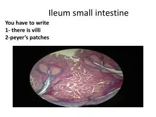
INTESTINAL EPITHELIAL CELLS. The panels below depict the bulk of this surface area expansion, showing villi, epithelial cells that cover the villi and the microvilli of the epithelial cells. Note in the middle panel, a light micrograph, that the microvilli are visible and look something like a brush. For this reason, the microvillus border of intestinal epithelial cells is referred to as the "brush border".
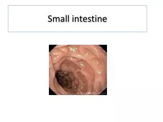
INTESTINAL EPITHELIAL CELLS. If examined closely, the lumenal surface of the small intestine appears similar to velvet due to it being covered by millions of small projections called villi which extend about 1 mm into the lumen. Villi are only the most obvious feature of the mucosa which houses a dynamic, self-renewing population of epithelial cells that includes secretory cells, endocrine cells and the mature absorptive epithelial cells which take up nutrients from the lumen and transport them into blood, fulfilling the basic function of the digestive system. Understanding how the small intestine functions requires looking at the structure of the mucosa in more detail. A light microscope view of epithelial cells from the small intestine. Magnification x 1000.
EPITHELIAL CELL DYNAMICS. Villi are projections into the lumen covered predominantly with mature, absorptive enterocytes, along with occasional mucus-secreting goblet cells. These cells live only for a few days, die and are shed into the lumen to become part of the ingesta to be digested and absorbed. That's right, we're all really cannibals. Crypts (of Lieberkuhn) are moat-like invaginations of the epithelium around the villi, and are lined largely with younger epithelial cells which are involved primarily in secretion. Toward the base of the crypts are stem cells, which continually divide and provide the source of all the epithelial cells in the crypts and on the villi. • The mucosa of small intestinal mucosa is arranged into two fundamental structures:
EPITHELIAL CELL DYNAMICS. • Coordinated contractions of smooth muscle participate in several ways to facilitate digestion and absorption in the small intestine: • foodstuffs are mixed with digestive enzymes from the pancreas and bile salts from the biliary system • nutrient molecules in the lumen are constantly dispersed, allowing them to contact the epithelium where enzymatic digestion is completed and absorption occurs • chyme is moved down the digestive tube, making way for the next load and also eliminating undigestable, perhaps toxic substances • In most animals, the small intestine cycles through two states: • Following a meal, when the lumen of the small intestine contains chyme, two types of motility predominate: segmentation contractions chop, mix and roll the chyme and peristalsis slowly propels it toward the large intestine. • The interdigestive state is seen between meals, when the lumen is largely devoid of contents. During such times, so-called housekeeping contractions propagate from the stomach through the entire small intestine, sweeping it clear of debris. This complex pattern of motility is the cause of "growling".
EPITHELIAL CELL DYNAMICS. The tight junctions between cells are impermeable to large organic molecules from the diet (e.g. amino acids and glucose). Those types of molecules are transported exclusively by the transcellular route, and only because the plasma membrane of the absorptive enterocytes is equipped with transporter molecules that facilitate entry into and out of the cells. An electron microscope view of epithelial cells from the small intestine. Magnification x 2000.
EPITHELIAL CELL DYNAMICS. It is important to recognize that the epithelium of the gut is not a monotonous sheet of functionally identical cells. As ingesta travels through the intestine, it is sequentially exposed to regions having epithelia with very different characteristics. This diversity in function results from differences in phenotype of the enterocytes - that is, the number and type of transporter molecules they express in their plasma membrane and the structure of the tight junctions they form. Even within a given segment there are major differences in the type of transport that occurs - for example, cells in the crypts transport very differently than cells on the tips of villi. Within the intestine, there is a proximal to distal gradient in osmotic permiability. As you proceed down the tube, the effective pore size through the epithelium decreases. This means that the duodenum is much more "leaky" to water than the ileum and the ileum more leaky than the colon. Do not interpret this to mean that as you go down the tube, the ability to absorb water decreases! It means that water flows across the epithelium more "freely" in the proximal compared to distal gut because the effective pore size is larger. The distal intestine actually can absorb water better than the proximal gut. The observed differences in permiability to water across the epithelium is due almost entirely to differences in conductivity across the paracellular path - the takehome message is that tight junctions vary considerably in "tightness" along the length of the gut.
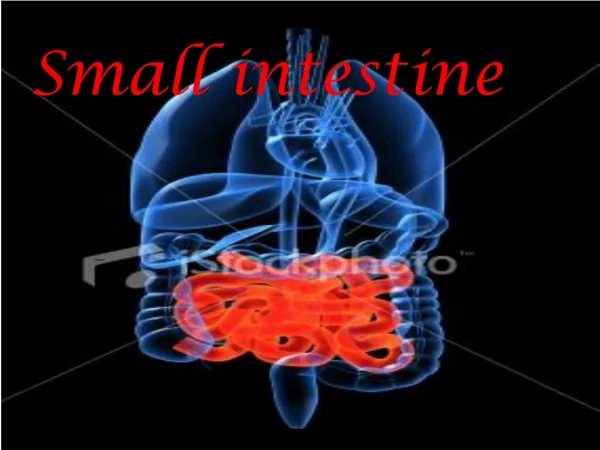
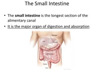
ORGANS. • An organ is a group of physically-linked different tissues working together to perform a specific physiological function. A picture of the small intestine.
TISSUES. A tissue is a group of similar cells performing a particular function. • Simple tissues are composed of one type of cell, while; • Compound tissues are composed of more than one type of cell.