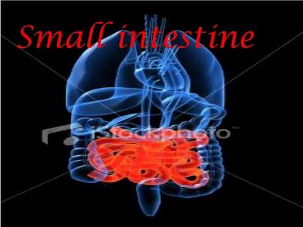
SMALL INTESTINE
SMALL INTESTINE . Dr IramTassaduq. SMALL INTESTINE. The small intestine is divided duodenum jejunum ileum. The duodenum is the first section of the small intestine and has a thicker layer of tissue than the other areas of the small intestine.


SMALL INTESTINE
E N D
Presentation Transcript
SMALL INTESTINE Dr IramTassaduq
SMALL INTESTINE • The small intestine is divided • duodenum • jejunum • ileum.
The duodenum is the first section of the small intestine and has a thicker layer of tissue than the other areas of the small intestine. It neutralizes stomach acids and breaks down carbohydrates and fats. The duodenum is about 2 feet long. THE DUODENUM
The jejunum is the main section of the small intestine. It covers about 15 feet and is responsible for the absorption of almost all nutrients except water. JEJUNUM
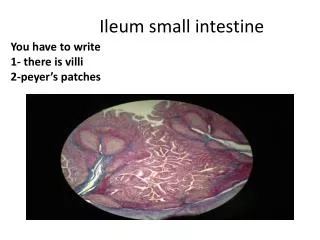
ILEUM The ileum is the last section of the small intestine and spans about 6 feet. Its function is to absorb water and vitamins.
PLICAE CIRCULARES • (valves of Kerkering) are macroscopically visible, crescent-shaped folds of the mucosa and submucosa. • permanent structures, i.e. their presence does not depend on the state of distension of the small intestine. • are absent from the first few centimetres of the duodenum and the distal part of the ileum. • well developed in the jejunum. • increase the surface area of the mucosa
INTESTINAL VILLI • The entire intestinal mucosa forms intestinal villi (about one mm long), which increase the surface area by a factor of ten. The surface of the villi is formed by a simple columnar epithelium.
GOBLET CELLS • The apical end of each goblet cell is occupied by a large mass of mucus, which compresses adjacent cells. • The nucleus toward the basal end of the cell. • Attached by junctional complexes (evidenced in light microscopy as the "terminal bar") to adjacent absorptive cells .
PANETH CELLS • Paneth cells are secretory epithelial cells located at the ends of intestinal crypts. The function for these cells is secretion of anti-bacterial proteins into the crypt lumen, thereby providing protection for the stem cells which line the crypt walls.
PANETH CELLS • Paneth cells have typical serous-secretory appearance, with basophilic basal cytoplasm (containing protein-synthetic rough endoplasmic reticulum) and apical secretory vesicles (zymogen granules).
ENTEROENDOCRINE CELLS • Concentrated in lower portion of intestinal gland • Produce a lot of peptide hormones