Download

1 / 31
310 likes | 328 Views
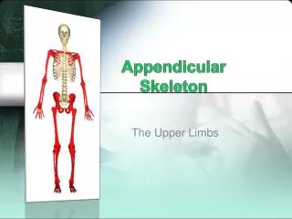
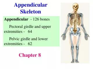
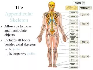
This study guide provides detailed views and descriptions of the bones in the appendicular skeleton, including the scapula, humerus, ulna, radius, carpal bones, and pelvic girdle. It also covers the bones of the hip and thigh, as well as the bones of the lower leg and foot.

E N D
The Appendicular Skeleton Study Guide
Superior border Suprascapularnotch Acromion Superior angle Coracoidprocess Glenoidcavity Subscapularfossa Lateral border Medial border Inferior angle Right scapula, anterior aspect
Coracoid process Suprascapular notch Superior angle Acromion Supraspinousfossa Glenoidcavityat lateralangle Spine Infraspinousfossa Lateral border Medial border Right scapula, posterior aspect
Greatertubercle Head ofhumerus Lessertubercle Deltoid tuberosity Coronoidfossa Medial epicondyle Capitulum Trochlea Anterior view
Head ofhumerus Greatertubercle Deltoidtuberosity Olecranonfossa Medial epicondyle Lateralepicondyle Trochlea Posterior view
Olecranon Troclearnotch Head Neck Coronoid process Interosseousmembrane Ulna Radius Head of ulna Ulnar styloidprocess Radial styloidprocess Anterior view
Olecranon Trochlear notch View Coronoid process Radial notch Proximal portion of ulna, lateral view
View Proximal portion of ulna, lateral view
The humerus of the right arm and detailed views of articulation at the elbow. Coronoidfossa Humerus Medialepicondyle Capitulum Trochlea Head ofradius Coronoidprocess ofulna Radialtuberosity Radial notch Radius Ulna Anterior view at the elbow region
The humerus of the right arm and detailed views of articulation at the elbow. Anterior view at the elbow region
Carpal bones thumb► “Sally left the party thumb►to take Carmen home” IV III II V Hamate I Capitate Trapezium Trapezoid Pisiform Scaphoid Triquetrum Lunate Ulna Radius Anterior view of right hand
Carpal bones IV III II V I Anterior view of right hand
Iliac crest Iliac fossa Anterior superioriliac spine Ilium Coxal bone Anteriorinferior iliacspine (os coxaeor hip bone) Sacrum Coccyx Pubis Acetabulum Ischium Pubic symphysis Pelvic girdle Pubic arch
Iliac fossa Sacrum Coccyx Pelvic girdle Pubic arch
Ilium Ala Iliac crest Anteriorsuperioriliac spine Posteriorsuperioriliac spine Anterior inferioriliac spine Posterior inferioriliac spine Acetabulum Greater sciaticnotch Ischial spine Lesser sciaticnotch Pubis Ischium Ischialtuberosity Ilium Ischium Pubis Lateral view, right hip bone
Ala Ilium Ischium Pubis Lateral view, right hip bone
Foveacapitis Neck Greatertrochanter Head Lesser trochanter Gluteal tuberosity Lateralcondyle Lateralepicondyle Medial condyle Lateralepicondyle Medialepicondyle Anterior view Posterior view Femur (thigh bone)
Anterior view Posterior view Femur (thigh bone)
Lateralcondyle Medialcondyle Head Tibialtuberosity Interosseousmembrane Fibula Tibia Medialmalleolus Lateralmalleolus Anterior view
Medialcuneiform Intermediatecuneiform Lateralcuneiform Navicular Cuboid Talus Calcaneus Superior view
Navicular Intermediate cuneiform Lateral cuneiform Talus Calcaneus Fifthmetatarsal Cuboid Lateral view
Talus Intermediatecuneiform Navicular Firstmetatarsal Medialcuneiform Calcaneus Medial view