Chapter 24
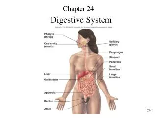
Chapter 24. Digestive System. Digestive System Anatomy. Digestive tract: also called alimentary tract or canal GI tract : technically refers to stomach and intestines Accessory organs Primarily glands, secrete fluids into tract Regions

Chapter 24
E N D
Presentation Transcript
Chapter 24 Digestive System
Digestive System Anatomy • Digestive tract: also called alimentary tract or canal • GI tract: technically refers to stomach and intestines • Accessory organs • Primarily glands, secrete fluids into tract • Regions • Mouth or oral cavity with salivary glands and tonsils • Pharynx (throat) with tubular mucous glands • Esophagus with tubular mucous glands • Stomach with many different kinds of glands that are tubular • Small intestine (duodenum, ileum, jejunum) with liver, gallbladder and pancreas as major accessory organs • Large intestine including cecum, colon, rectum and anal canal with mucous glands • Anus
Functions • Ingestion: introduction of food into stomach • Mastication: chewing. Chemical digestion requires large surface area so breaking down large particles mechanically facilitates chemical digestion. • 3.Propulsion (movement of food-----24-36 hours oral end to anal end) • Deglutition: swallowing (oral cavity -> esophagus) (bolus = mass of food or liquid) • Peristalsis: moves material through digestive tract . A wave of circular smooth muscle relaxation moves ahead of the bolus of food or chyme allowing the digestive tract to expand. Then a wave of contraction of the circular smooth muscles behind the bolus of food or chyme (ingested food & stomach secretions) propels it through the digestive tract. • Mass movements in large intestine (contractions that extend for larger parts of digestive tract)
4. Mixing: Segmental contractions ( mixing contractions that occur in small intestine. -Some contractions do not propel food from one end of digestive tract to the other but, rather, move it back & forth within digestive tract to mix it with digestive secretions & help break it into smaller pieces)
Functions, cont. 5.Secretion: lubricate, liquefy, digest • Mucus: secreted along entire digestive tract, lubricates food and lining, coats lining and protects from mechanical digestion, from acid and from digestive enzymes. • Water: liquefaction makes food easier to digest and absorb • Bile: emulsifies fats • Enzymes: chemical digestion 6.Digestion: Mechanical and chemical 7.Absorption: Movement from tract into circulation or lymph 8.Elimination: Waste products removed from body; feces. Defecation
Digestive Tract Histology: The Tunics • Mucosa. Innermost layer, consisting of mucous epithelium (stratified squamous in mouth, oropharynx, esophagus and anal canal), simple columnar epithelium in the rest of the tract. • Loose connective tissue: lamina propria • Muscularis mucosae: smooth muscle • Submucosa. Thick C.T. layer with nerves, blood vessels, small glands. Parasympathetic submucosal plexus.
Digestive Tract Histology: The Tunics • Muscularis: 2 or 3 layers of smooth muscle, two of which are circular and longitudinal. Exception: esophagus where the upper 1/3 is striated & stomach. This layer also contains the myenteric plexus. The myenteric and submucosal plexi together are called the enteric or intramural plexus. Important in control of movement and secretion • Serosa or adventitia: Connective tissue. Where serosa is present, called visceral peritoneum. Where adventitia is present, connective tissue blends with connective tissue of surrounding structures
Nervous regulation Local: enteric nervous system Types of neurons: sensory, motor, interneurons Coordinates peristalsis and regulates local reflexes General: coordination with the CNS. May initiate reflexes because of sight, smell, or taste of food. Parasympathetic primarily (through vagas nerve). Sympathetic input inhibits muscle contraction, secretion, and decrease of blood flow to the digestive tract. Chemical regulation Production of hormones to be discussed later Gastrin, secretin Production of paracrine chemicals like histamine Help local reflexes in ENS control the conditions of the internal environment of the digestive tract such as pH levels Digestive System Regulation
Peritoneum and Mesenteries • Peritoneum • Visceral: Covers organs • Parietal: Covers interior surface of body wall • Retroperitoneal: Certain organs covered by peritoneum on only one surface and are considered behind the peritoneum; (lie against abdominal wall) e.g., kidneys, pancreas, duodenum • Mesenteries: two layers of peritoneum with thin layer of loose C.T. between • Routes by which vessels and nerves pass from body wall to organs • Greater omentum: connects greater curvature of the stomach to the transverse colon (extends inferiorly from stomach over surface of small intestine). • Lesser omentum: connects lesser curvature of the stomach and the proximal part of the duodenum to the liver and diaphragm. • Transverse mesocolon, sigmoid mesocolon, mesoappendix (mesentery refers to serous membranes attached to abdominal organs). • Ligaments • Coronary: between liver and diaphragm • Falciform: between liver and anterior abdominal wall
Oral Cavity • Bounded by lips anteriorly, fauces (opening into pharynx) posteriorly • Vestibule: space between lip/cheeks and alveolar processes with teeth • Oral cavity proper: medial to alveolar processes • Lined with moist stratified squamous epithelium
Lips and Cheeks • Both structures important in mastication and speech • Lips (labia): orbicularis oris muscle within. Keratinized stratified squamous exterior is thin and color of blood in dermis gives a red/pink color. • Labial frenula (mucous folds) extend from alveolar processes of maxilla and mandible to the upper and lower lips, respectively. • Many facial muscles act to move lips • Cheeks: lateral walls of oral cavity • Buccinator muscle • Buccal fat pad
Palate and Palatine Tonsils • Palate • Hard palate: anterior, supported by maxilla and palatine bone • Soft palate: posterior, consists of skeletal muscle and connective tissue • Uvula: projects from posterior of soft palate • Palatine tonsils: lateral walls of fauces
Tongue • Muscular with free anterior surface and attached posterior surface. Covered with moist stratified squamous epithelium. • Intrinsic muscles: change shape • Extrinsic muscles: protrude or retract tongue, move side to side • Lingual frenulum attaches tongue inferiorly to floor of oral cavity • Terminal sulcus: groove divides tongue into anterior 2/3; posterior 1/3 • Anterior part: papillae, some of which have taste buds • Posterior part: no papillae and a few scattered taste buds. Lymphoid tissue embedded in posterior surface: lingual tonsil • Moves food in mouth, participates in speech and swallowing
Teeth • Two sets • Primary, deciduous, milk: Lost during childhood • Permanent or secondary: Adult (32) • Types • Incisors, canines, premolars and molars
Teeth • Involved in mastication and speech • Anatomic crown: enamel-covered part of tooth; clinical crown is section of tooth above gum line • Neck: enameled part of tooth below gum line • Enamel: outermost layer of anatomical crown. Non-living; acellular. Protective. • Dentin: living, cellular, calcified tissue. In the root, dentin is covered by cellular bone-like structure that helps hold tooth in the socket. • Pulp cavity filled with blood vessels, nerves, and connective tissue • Periodontal ligaments: hold tooth in socket. • Gingiva: dense, fibrous C.T. covered by stratified squamous epithelium.
Mastication • Chewing: incisors and canines bite or cut off food; molar-type teeth grind food • Muscles involved: masseter, temporalis, medial and lateral pterygoids. • Elevate mandible (close jaw): temporalis, masseter, medial pterygoids • Depress mandible (open jaw): lateral pterygoids • Protraction (moving in anterior direction) and lateral and medial excursion (lateral = moves mandible to either right or left of midline--------medial = returns mandible to neutral position): pterygoids and masseter • Retraction (moves structure back to anatomical position)- temporalis • Mastication reflex: medulla oblongata, but descending pathways from cerebrum provide conscious control. Controls basic movements involved in chewing
Salivary Glands • Three pairs of multicellular glands • Parotid: largest. Serous. Just anterior to the ear. Parotid duct crosses over masseter, penetrates buccinator, and enters the oral cavity adjacent to the 2nd upper molar • Submandibular: mixed, but more serous than mucous. Posterior half of inferior border of mandible. Duct enters oral cavity on either side of lingual frenulum • Sublingual: smallest. Mixed, but primarily mucous. Each has 10-12 ducts that enter the floor of the oral cavity. • Lingual glands. Small, coiled tubular glands on surface of tongue.
Saliva • Compound alveolar salivary glands. Produce saliva • Prevents bacterial infection • Lubrication • Contains salivary amylase that breaks down starch into disaccharides maltose and isomaltose (gives starch sweet taste in mouth). • Helps to form bolus for swallowing • Parasympathetic input causes salivary production
Pharynx Posterior walls of oropharynx and laryngopharynx contains group of muscles called pharyngeal constrictors that contribute to swallowing Esophagus Transports food from pharynx to stomach Passes through esophageal hiatus (opening) of diaphragm and ends at stomach Hiatal hernia: widening of hiatus (causes ulcers, acid reflux) Sphincters Upper. Striated Lower. Smooth Mucosa is moist stratified squamous epithelium. Produces thick layer of mucus. Pharynx and Esophagus
Swallowing (Deglutition) • Three phases • Voluntary: bolus of food moved by tongue from oral cavity to pharynx. • Pharyngeal: reflex. Controlled by swallowing center in medulla oblongata. Soft palate elevates, upper esophageal sphincter relaxes, elevated pharynx opens the esophagus, food pushed into esophagus by pharyngeal constrictors’ successive contraction from superior to inferior. Epiglottis is tipped posteriorly due to pressure of the bolus, larynx elevated to prevent food from passing into larynx. • Esophageal: reflex. Stretching of esophagus causes enteric NS to initiate peristalsis of muscles in the esophagus.
Stomach Anatomy • Greater and lesser curvatures: attachment sites for omenta • Sphincters • Cardiac (lower esophageal) • Pyloric • Openings • Gastroesophageal (cardiac): to esophagus • Pyloric: to duodenum • Parts • Cardiac • Fundus • Body • Pyloric: antrum and canal
Stomach Histology • Rugae: folds in stomach when empty. Mucosa and submucosa. • Layers • Serosa or visceral peritoneum • Muscularis: three layers • Outer longitudinal • Middle circular • Inner oblique (Having a slanting or sloping direction) • Submucosa • Mucosa
Stomach Histology • Gastric pits: openings for gastric glands. Lined with simple columnar epithelium • Cells of gastric pits • Surface mucus: mucus that protects stomach lining from acid and digestive enzymes • Mucous neck: mucus • Parietal: hydrochloric acid and intrinsic factor • Chief: pepsinogen • Endocrine: regulatory hormones • Enterochromaffin-like cells: secretes histamine that stimulates acid secretion • Gastrin-containing cells: secrete gastrin (a hormone that stimulates acid secretion) • Somatostatin-containing cells: secrete somatostatin that inhibits gastrin and insulin secretion
Secretions of the Stomach • Chyme: ingested food plus stomach secretions • Mucus: surface and neck mucous cells • Viscous and alkaline • Protects from acidic chyme and enzyme pepsin • Irritation of stomach mucosa causes greater mucus • Intrinsic factor: parietal cells. Binds with vitamin B12 and helps it to be absorbed in the ileum. B12 necessary for DNA synthesis and RBC production (lack of B12 absorption leads to pernicious anemia) • HCl: parietal cells • Kills bacteria (found in ingested food) • Stops carbohydrate digestion by inactivating salivary amylase • Denatures proteins • Helps convert pepsinogen to pepsin (optimal activity at pH 3 or less) • Pepsinogen: packaged in zymogen granules released by exocytosis. Pepsin catalyzes breaking of covalent bonds in proteins (breaks them into smaller peptide chains)
Cephalic Phase • The taste or smell of food, tactile sensations of food in the mouth, or even thoughts of food stimulate the medulla oblongata. • Parasympathetic action potentials are carried by the vagus nerves to the stomach, where enteric plexusneurons are activated. • Postganglionic neurons stimulate secretion by parietal and chief cells (HCl and pepsin) and stimulate the secretion of the hormone gastrin and histamine. • Gastrin is carried through the circulation back to the stomach where it and histamine stimulate further secretion of HCl and pepsin.
GastricPhase • Distention of the stomach activates a parasympathetic reflex. Action potentials are carried by the vagus nerves tothe medulla oblongata. • Medulla oblongata stimulates further secretions of the stomach. • Distention also stimulates local reflexes that amplify stomach secretions.
IntestinalPhase • Chyme in the duodenum with a pH less than 2 or containing lipids inhibits gastric secretions by three mechanisms • Sensory input to the medulla from the duodenum inhibits the motor input from the medulla to the stomach. Stops secretion of pepsin and HCl. • Local reflexes inhibit gastric secretion • Secretin, and cholecystokininproduced by the duodenum decrease gastric secretions in the stomach.
Movements in Stomach • Combination of mixing waves (80%) and peristaltic waves (20%) • Both esophageal and pyloric sphincters are closed.
Small Intestine • Site of greatest amount of digestion and absorption of nutrients and water • Divisions • Duodenum- first 25 cm beyond the pyloric sphincter. • Jejunum- 2.5 m • Ileum- 3.5 m. Peyer’s patches or lymph nodules
Duodenum • Curves to the left; head of pancreas in the curve • Major and minor duodenal papillae: openings to ducts from liver and/or pancreas.
Modifications to Increase Surface Area • Increase surface area 600 fold • Plicae circulares (circular folds) • Villi that contain capillaries and lacteals. Folds of the mucosa • Microvilli: folds of cell membranes of absorptive cells
Mucosa and Submucosa of the Duodenum • Cells and glands of the mucosa • Absorptive cells: cells with microvilli, produce digestive enzymes and absorb digested food • Goblet cells: produce protective mucus • Endocrine cells: produce regulatory hormones (Secretin, and cholecystokinin) • Granular cells (paneth cells): may help protect from bacteria (contain lysozymes) • Intestinal glands (crypts of Lieberkühn): tubular glands in mucosa at bases of villi [secrete sucrase ,maltase, trypsin, chymotrypsin, and pepsin (endopeptidases and exopeptidases) ] • Duodenal glands (Brunner’s glands): tubular mucous glands of the submucosa. Open into intestinal glands [produce a mucus-rich alkaline secretion (containing bicarbonate)
Jejunum and Ileum • Gradual decrease in diameter, thickness of intestinal wall, number of circular fold, and number of villi the farther away from the stomach • Major site of nutrient absorption • Peyer’s patches: lymphatic nodules numerous in mucosa and submucosa • Ileocecal junction: where ileum meets large intestine. Ileocecal sphincter (ring of smooth muscle) and ileocecal valve (one-way valve)
Small Intestine Secretions • Fluid primarily composed of water, electrolytes and mucus. • Mucus • Protects against digestive enzymes and stomach acids • Digestive enzymes: bound to the membranes of the absorptive cells • Disaccharidases: Break down disaccharides to monosaccharides • Peptidases: Hydrolyze peptide bonds • Nucleases: Break down nucleic acids • Duodenal glands • Stimulated by vagus nerve, secretin, chemical or tactile irritation of duodenal mucosa
Movement in Small Intestine • Mixing and propulsion over short distances • Segmental contractions mix • Peristalsis propels • Ileocecal sphincter remains slightly contracted until peristaltic waves reach it; it relaxes, allowing chyme to move into cecum • Cecal distention causes local reflex and ileocecal valve constricts • Prevents more chyme from entering cecum • Increases digestion and absorption in small intestine by slowing progress of chyme • Prevents backflow
Liver • Lobes • Major: Left and right • Minor: Caudate and quadrate • Porta: on inferior surface. Vessels, ducts, nerves, exit/enter liver • Hepatic portal vein, hepatic artery, hepatic nerve plexus enter • Lymphatic vessels, two hepatic ducts exit • Ducts • Right and lefthepatics (which transport bile out of liver) unite to form • Common hepatic • Cystic: from gallbladder • Common bile: union of cystic duct and common hepatic duct (common bile joins the pancreatic duct at the hepatopancreatic ampulla------ampulla empties into duodenum at major duodenum papilla)
Histology of the Liver • Connective tissue septa branch from the porta into the interior • Divides liver into lobules • Nerves, vessels and ducts follow the septa • Lobules: portal triad at each corner • Three vessels: hepatic portal vein, hepatic artery, hepatic duct • Central vein in center of lobule • Central veins unite to form hepatic veins that exit liver and empty into inferior vena cava
Liver Histology • Hepatic cords: radiate out from central vein. Composed of hepatocytes • Hepatic sinusoids: between cords, lined with endothelial cells and hepatic phagocytic (Kupffer) cells • Bile canaliculus: between cells within cords • Hepatocyte functions • Bile production • Storage • Interconversion of nutrients • Detoxification • Phagocytosis • Synthesis of blood components
Functions of the Liver • Bile production: 600-1000 mL/day. Bile salts, bilirubin (bile pigment that results from breakdown of hemoglobin), cholesterol, fats, fat-soluble hormones, lecithin • Neutralizes and dilutes stomach acid (neutralizes chyme so that pancreatic enzymes can function) • Bile salts emulsify fats. Most are reabsorbed in the ileum. (90% bile salts reabsorbed in the ileum & carried back to liver) • Secretin (from the duodenum) stimulates bile secretions, increasing water and bicarbonate ion content of the bile • Storage • Glycogen, fat, vitamins (A, B12, D, E, and K), copper and iron. Hepatic portal blood comes to liver from small intestine (nutrients are stored and secreted back into circulation when needed) • Synthesis • Blood proteins: Albumins, fibrinogen, globulins, heparin, clotting factors (liver produces its own new compounds)
Functions of the Liver • Nutrient interconversion • Amino acids to energy producing compounds (ex: person on a excessively high protein diet and low fat & carb diet----------an oversupply of amino acids & an undersupply of lipids & carbs are delivered to the liver. The hepatocytes break down the amino acids and cycle them through metabolic pathways so they can be used to produce adenosine triphosphate, lipids, and glucose) • Hydroxylation of vitamin D. Vitamin D then travels to kidney where it is hydroxylated again into its active form • Hepatocytes also transform substances that cannot be used by most cells into usable sunstances. (ex: ingested fats combined with choline {nutrient in B vitamin family} & phosphorous in liver to produce phospholipids, which are imp. for cell membranes) • Detoxification • Hepatocytes remove ammonia (by-product of amino acid metabolism) which is toxic & not readily removed by kidneys. Hepatocytes convert it to urea which is less toxic and easily eliminated by kidneys. • Phagocytosis • Kupffer cells phagocytize worn-out and dying red and white blood cells, some bacteria
Gallbladder • Sac lined with mucosa folded into rugae, inner muscularis, outer serosa • Bile arrives constantly from liver is stored and concentrated • Stimulated by cholecystokinin (from the intestine) and vagal stimulation • Bile exits through cystic duct then into common bile duct • Gallstones: precipitated cholesterol (occurs when excess cholesterol in bile due to high-cholesterol diet and not enough bile salts to keep it in solution) • Can block cystic duct • If gallstone moves far down the duct, it can block pancreatic duct, resulting in pancreatitis. • Can occur because of drastic dieting(as the body metabolizes fat during prolonged fasting and rapid weight loss—such as “crash diets”—the liver secretes extra cholesterol into bile, which can cause gallstones.)
Pancreas • Pancreas both endocrine and exocrine • Head, body and tail • Endocrine: pancreatic islets. Produce insulin, glucagon, and somatostatin • Exocrine: groups acini (grape-like cluster) form lobules separated by septa. • Intercalated ducts lead to intralobular ducts lead to interlobular ducts lead to the pancreatic duct. • Pancreatic duct joins common bile duct and enters duodenum at the hepatopancreatic ampulla controlled by the hepatopancreatic ampullar sphincter
Pancreatic Secretions: Pancreatic Juice • Aqueous. Produced by columnar epithelium lining smaller ducts. Na+, K+, HCO3-, water. Bicarbonate lowers pH inhibiting pepsin and providing proper pH for enzymes • Enzymatic portion: (without the enzymes produced by pancreas, lipids, proteins, & carbs not adequately digested) • Trypsinogen- active form is trypsin--------proteolytic enzyme • Chymotrypsinogen- active form is chymotrypsin--------proteolytic enzyme • Procarboxypeptidase- active form is carboxypeptidase-------proteolytic enzyme • Pancreatic amylase- continues digestion of starch. • Pancreatic lipases- lipid digesting enzyme • Deoxyribonucleases and ribonucleases- reduce DNA & RNA to their nucleotide • Interaction of duodenal and pancreatic enzymes • Enterokinase is a proteolytic enzyme from the duodenal mucosa and it activates trypsinogen to trypsin. • Trypsin activates chymotrypsinogen to chymotrypsin. • Trypsin activates procarboxypeptidase to carboxypeptidase.