Download
1 / 37
370 likes | 460 Views
The Musculoskeletal System. MEDL 2350. Quiz. xanthoerythemoectooculoscopy Salpingophthalmosclerodermatitioscope Blepharopepicosclerotomy Gluconeothymomotomically Pancreatectomicaoloma Only kidding, ha, ha!. So, you want extra credit, huh!. MEDL 2350 Extra Credit. TEST on May 5, 2003

E N D
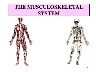
The Musculoskeletal System MEDL 2350
Quiz • xanthoerythemoectooculoscopy • Salpingophthalmosclerodermatitioscope • Blepharopepicosclerotomy • Gluconeothymomotomically • Pancreatectomicaoloma • Only kidding, ha, ha!
MEDL 2350 Extra Credit TEST on May 5, 2003 First half of class Topics: EVERYTHING Format: Oral Counts: Test Grade
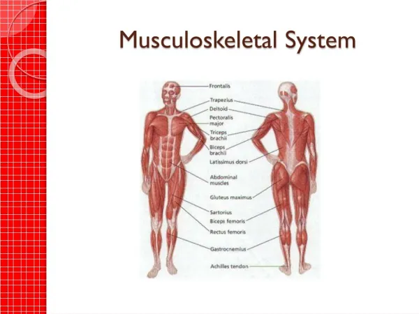
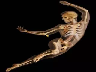
General • Comprised of bones, joints, and muscles. • Bones form a support system to provide structure and protection. • Bones store minerals, especially calcium and phosphorus. • Bones also make blood cells in the marrow. • The place where two bones meet is known as a joint. • Joints move due to the pull of muscles.
Combining Forms Crani/o cranium (skull bones) craniotomy Stern/o sternum (breastbone) sternocostal
Cost/o ribs subcostal (below the ribs) Spondyl/o vertebrae (backbone) Vertebr/o spondylitis vertebral
Humer/o humerus (upper arm) Carp/o carpus (wrist) carpoptosis (dropping of the wrist)
Metacarp/o metacarpus (hand) metacarpectomy excision of a metacarpal Phalang/o finger/toe Pelv/i pelvis (hip) *note the exception to the combining vowel. pelvimetry (process of measuring the hips)
Femor/o femur (thigh) Patell/o patella (kneecap) patellopexy (fixation) Tibi/o tibia (shin) Fibul/o fibula (smaller lower leg bone) Calcane/o calcaneus (heel) Tars/o tarsals metatars/o metatarsals
Ankyl/o stiff, bent, crooked Arthr/o joint cervic/o neck Lamin/o lamina (part of the verterbral arch) Myel/o spinal cord, bone marrow Orth/o straight Oste/o bone
The combining form oste/o refers to a bone. Therefore, osteodynia refers to painful bones. Osteocytes are cells that make up bones • The combining form still holds true regardless of the shape of the bone in question.
Shapes of Bones • Long (cylindrical) • Flat • Irregular • short
Milk is source of vitamin D. A deficiency of this vitamin results in Osteomalacia, or a softening and weakening of the bones. • This is different than Osteoporosis, which is a deficiency of bone mineral. • The term osteogenesis refers to producing or forming new bone.
Anatomy of a Long Bone • Shaft: Diaphysis • Epiphysis: either end, the growing part in length. • Bone Marrow: soft tissue that fills the medullary cavity in the center of a long bone. This is where blood is formed and fat is stored. • Periosteum: thin sheet of tissue covering the bone. This part of the bone is responsible for growth in the diameter.
X-rays or radiographs are used in examining the structure of bones, in situ (within the body). • The combining form for x-ray or radiation is radi/o. • The combining forms roentgen/o also refers to x-rays. This is after the person who discovered x-rays, Wilhelm Roentgen.
mening/o/myel/o/cele herniation Meninges- covering of the brain and spinal cord Spinal cord
Spina bifida usually occurs in the lumbar region (lower back) of the spinal cord. Bifida refers to being in “2”.
Recall, Distal: means farthest from the trunk Proximal: means closest to the trunk Therefore, the distal epiphysis is located farther from the trunk than the proximal epiphysis.
Within the joints, each end of the joint (epiphysis) is lined with ARTICULAR CARTILAGE. • Articular means joint • Chondr/o refers to cartilage. • This articular cartilage provides cushioning, shock absorption, an area for the bones to glide easily over each other during movement.
A joint is a place where two bones meet. • Joints can be: Freely Moveable (shoulder) Slightly Moveable (vertebra) Non-moveable (skull) • Joints are lubricated with SYNOVIAL FLUID. This fluid is secreted within the SYNOVIAL MEMBRANE.
arthr/o/centesis joint Surgical puncture
A person with arthritis can suffer from: Pain in the joints - arthralgia or arthrodynia Inflammation of the joints - arthritis Disease of the bones and joints – osteoarthopathy
Combining Forms of Specific Bones Crani/o cranium skull Stern/o sternum breastbone cost/o costal ribs Vertebr/o veterbrae
Humer/o humerus carp/o carpus Metacarp/o metacarpal Phalang/o phalanges
Pelv/i pelvis hips Pelv/o Femor/o femur thigh patell/o patella kneecap Tibi/o tibia fibul/o fibula
Cephal/o is the combining form for the direction “close to the head” • Cephalodynia is pain in the head, or headache. • The prefix en- refers to “in”. When combined with cephal/o, when get encephal/o which translates into “in the head”. This the another term for brain.
Fractures • A fracture is a break in the continuity of a bone. They are defined according to the type and extent of the break. • A SIMPLE or CLOSED fracture is a broken bone with no external wound. The skin is intact. • A COMPLEX or OPEN fracture is one with an open wound. The skin is broken.
A GREENSTICK fracture is a bone that has been bent and partially broken. This is more common in children than adults. • A COMMINUTED fracture is one that is in many pieces.
The Vertebral Column • The vertebral column is made of many bones called vertebra. • The vertebral column is divided into 5 parts: Cervical 7 bones cervic/o Thoracic 12 bones thorac/o Lumbar 5 bones lumb/o Sacral 5 fused bones sacr/o Coccyx
Vertebrae are separated and cushioned from each other by INTERVERTEBRAL DISKS. • These disks are made of cartilage. • The first cervical vertebrae is called the ATLAS. The axis holds up the head. • The second cervical vertebrae is called the AXIS.
Muscular System Chondr/o cartilage Lumb/o lower back My/o muscle ten/o tendon Tend/o Tendin/o