Musculoskeletal System
2.55k likes | 3.66k Views
4. Musculoskeletal System. Multimedia Directory. Slide 81 Chiropractic Medicine Video Slide 94 Fracture Animation Slide 97 Osteoporosis Video Slide 105 Arthritis Video Slide 112 Arthroscopy Video Slide 140 Muscles Animation Slide 157 Humerus Adduction/Abduction Animation

Musculoskeletal System
E N D
Presentation Transcript
4 Musculoskeletal System
Multimedia Directory Slide 81 Chiropractic Medicine Video Slide 94 Fracture Animation Slide 97 Osteoporosis Video Slide 105 Arthritis Video Slide 112 Arthroscopy Video Slide 140 Muscles Animation Slide 157 Humerus Adduction/Abduction Animation Slide 159 Elbow Flexion/Extension Animation Slide 161 Ankle Dorsiflexion and Plantar Flexion Animation Slide 164 Ankle Inversion and Eversion Animation Slide 166 Elbow Pronation and Supination Animation Slide 168 Humerus Circumduction Animation Slide 169 Hand Opposition Animation Slide 170 Humerus Rotation Animation
Multimedia Directory Continued Slide 182 Muscle Atrophy Video Slide 189 Muscular Dystrophy Video Slide 191 Carpal Tunnel Video Slide 192 Carpal Tunnel Animation
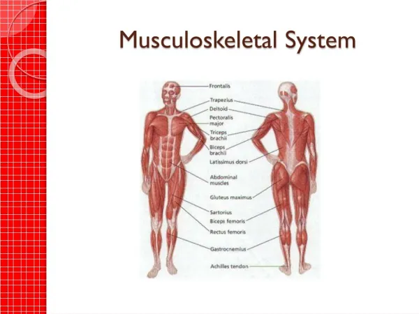
Skeletal System at a Glance • Functions of Skeletal System • Internal framework of body • Supports body • Protects internal organs • Point of attachment for muscles • Produces blood cells • Stores minerals
Skeletal System at a Glance • Organs of Skeletal System • Bones of the skeleton • Joints
Skeletal System Combining Forms • ankyl/o – stiff joint • arthr/o – joint • articul/o – joint • burs/o – sac • carp/o – wrist • cervic/o – neck • chondr/o – cartilage • clavicul/o – clavicle
Skeletal System Combining Forms • coccyg/o – coccyx • cortic/o – outer portion • cost/o – rib • crani/o – skull • femor/o – femur • fibul/o – fibula • humer/o – humerus • ili/o – ilium
Skeletal System Combining Forms • ischi/o – ischium • kyph/o – hump • lamin/o – lamina, part of vertebra • lord/o – bent backwards • lumb/o – low back, loin • mandibul/o – mandible • maxill/o – maxilla • medull/o – inner portion
Skeletal System Combining Forms • metacarp/o – metacarpals • metatars/o – metatarsals • myel/o – bone marrow • orth/o – straight • oste/o – bone • patell/o – patella • ped/o – foot, child • pelv/o – pelvis
Skeletal System Combining Forms • phalang/o – phalanges • pod/o – foot • prosthet/o – addition • pub/o – pubis • radi/o – radius, ray • sacr/o – sacrum • sarc/o – flesh • scapul/o – scapula
Skeletal System Combining Forms • scoli/o – crooked, bent • spin/o – spine • spondyl/o – vertebrae • stern/o – sternum • synovi/o – synovial membrane • synov/o – synovial membrane • tars/o – ankle • thorac/o – chest
Skeletal System Combining Forms • tibi/o – tibia • uln/o – ulna • vertebr/o – vertebra
Skeletal System Suffixes • –blast immature, embryonic • –clasia to break surgically • –desis stabilize, fuse • –listhesis slipping • –porosis porous
Anatomy and Physiology • Bones are body organs with blood supply, nerves, and lymphatic vessels • Bones are connected to each other to form skeleton • Framework for the body • 206 bones
Anatomy and Physiology • Red bone marrow within bones produces blood cells • Bones also: • Protect vital organs • Store minerals
Anatomy and Physiology • Joint • Place where two bones meet • Held together by ligaments • Gives flexibility to skeleton
Bones • Also called osseous tissue • One of hardest materials in body • Formed from gradual process before birth called ossification • Fetal skeleton is formed from a cartilage model
Bones • Flexible tissue is gradually replaced by osteoblasts (immature bone cells) • In adult bones osteoblasts mature into osteocytes • Formation of strong bones dependant on adequate supply of minerals
Long Bones Majority of bones in body Divided into: Diaphysis Epiphysis
Diaphysis • Central sht • Medullary cavity • Open canal within diaphysis • Contains yellow bone marrow • Mostly fat
Epiphysis Wide ends of long bone Distal epiphysis Proximal epiphysis Articular cartilage Covers epiphysis Prevents bone rubbing on bone
Periosteum Covers surface of bone not covered by articular cartilage Thin connective tissue membrane Contains numerous nerve and lymphatic vessels
Compact Bone Also called cortical bone Very dense and hard Outer layer of bone Found in both epiphysis and diaphysis
Cancellous Bone Also called spongy bone Found inside bone Has spaces containing red bone marrow Manufactures blood cells
Bony Processes • Projection from the surface of a bone • Rough processes provide place for muscle attachment • Smooth rounded processes articulate with another bone in a joint • Named for shape and location
The Skeleton • Skeleton has two divisions • Axial skeleton • Appendicular skeleton
Axial Skeleton • Includes bones in: • Head • Neck • Spine • Chest • Trunk
The Skull • Is divided into two parts • Cranium • Facial bones • Protects brain, eyes, ears, nasal cavity, and oral cavity • Attachment for muscles of chewing and turning the head
Cranium • Frontal – 1 • Forehead • Parietal – 2 • Upper sides and roof of skull • Temporal – 2 • Sides & base of skull
Cranium • Ethmoid – 1 • Part of eye orbit, nose, & floor of skull • Sphenoid – 1 • Part of floor of skull • Occipital – 1 • Back & base of skull
Facial Bones • Mandible – 1 • Lower jawbone • Maxilla – 1 • Upper jawbone • Zygomatic – 2 • Cheek bones • Vomer – 1 • Part of nasal septum
Facial Bones • Palatine – 1 • Hard palate and floor of nose • Nasal – 2 • Part of nasal septum and bridge of nose • Lacrimal – 2 • Inner corner of eye
Hyoid Bone • Single U-shaped bone • In neck between mandible and larynx • Attachment point for swallowing and speech muscles
The Trunk • Vertebral column • Sternum • Rib cage
The Vertebral Column • Divided into five sections • Cervical • Thoracic • Lumbar • Sacrum • Coccyx
The Vertebral Column • Cervical • 7 vertebrae of neck • Thoracic • 12 vertebrae of chest • Lumbar • 5 vertebrae of low back
The Vertebral Column • Sacrum • 5 fused vertebrae at base of spine • Coccyx • 3–5 small vertebrae attached to sacrum
The Rib Cage • 12 pairs of ribs • Attached to vertebral column at back • Provides support for organs, such as heart and lungs
The Rib Cage • True ribs • 10 pairs attached to sternum in front • Floating ribs • Inferior 2 pairs • No attachment in front