Download
1 / 52
590 likes | 1.01k Views
Chapter 27. The Musculoskeletal System. Learning Objectives (1 of 2). Name common congenital abnormalities of the skeletal system Describe three major types of arthritis, pathogenesis, clinical manifestations, and treatment Describe causes and effects of osteoporosis, and methods of treatment

E N D
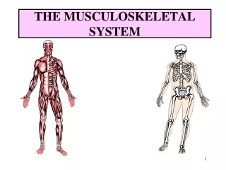
Chapter 27 The Musculoskeletal System
Learning Objectives (1 of 2) • Name common congenital abnormalities of the skeletal system • Describe three major types of arthritis, pathogenesis, clinical manifestations, and treatment • Describe causes and effects of osteoporosis, and methods of treatment • Describe structure and functions of intervertebral disks; clinical manifestations of herniated disk
Learning Objectives (2 of 2) • Describe pathogenesis, manifestations, and treatment of myasthenia gravis • Describe manifestations, complications, and treatment of scoliosis • Compare pathogenesis, common types, and clinical manifestations of muscular atrophy and dystrophy
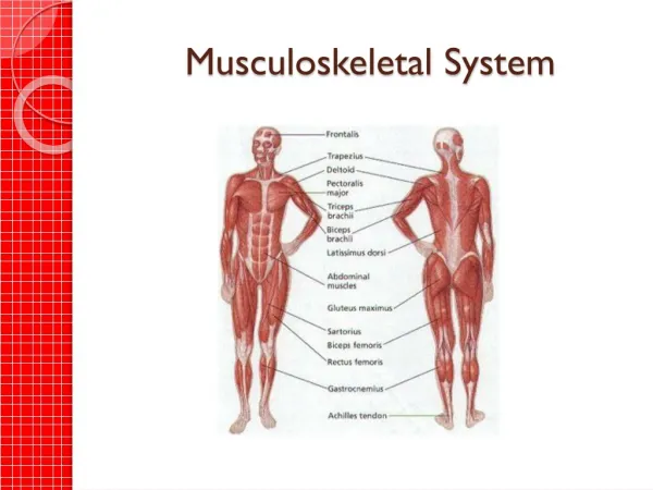
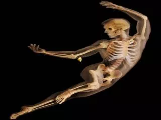
Skeletal System (1 of 2) • Skeleton: rigid supporting structure of the body • All bones have same basic structure • Cortex: outer layer of compact bone, the cortex • Trabeculae: inner spongy layer arranged in a loose meshed lattice of thin strands • Bone marrow: spaces between trabeculae consist of fat and blood-forming tissue • Bone: specialized type of connective tissue • Composed of a dense connective tissue framework impregnated with calcium phosphate salts • Continually broken down and reformed
Skeletal System (2 of 2) • Types of cells in bone • Osteoblasts • Osteocytes • Osteoclasts • Strength and thickness of bones depend on activity • Bones of skeleton are connected by joints • Types of joints • Fibrous joint • Cartilaginous joint • Synovial joint
Bone Formation • Intramembranous • Mesoderm transformed into osteoblasts that are converted into bone • Endochondral • Cartilage model converted into bone
Congenital Malformations (1 of 2) • Achondroplasia • Faulty endochondral bone formation • Impaired growth of extremities and formation of skull bones • Causes dwarfism with disproportionately short limbs • Osteogenesis imperfecta • Thin and delicate bones easily broken • May be born with multiple fractures • Malformation of fingers and toes • Extra digits or polydactyly • Easily removed • Fused digits more difficult to correct
Congenital Malformations (2 of 2) • Congenital clubfoot (talipes) • Multifactorial inheritance • Most common type: talipes equinavarus • Treatment: manipulation and casts • Congenital dislocation of the hip • Multifactorial inheritance • More common in females • Shallow acetabulum causes femoral head to be displaced out of socket • Breech position favors development • Treatment: manipulation and casts
Common malformations of fingers. Failure of separation of fingers.
Rheumatoid Arthritis (RA) • Systemic disease affecting connective tissues throughout the body, specially the joints • Produces chronic inflammation and thickening of synovial membrane • Classified as an autoimmune disease • Rheumatoid factor: autoantibody in blood and synovial tissues; produced by B lymphocytes directed against individual’s own gamma globulin • Encountered most frequently in young men and middle-aged women • Usually affects small joints of hands and feet • Dislocation from joint instability
Photomicrograph of rheumatoid arthritis illustrating destruction of articular cartilage by inflammatory reaction.
Photomicrograph of rheumatoid arthritis. Chronic inflammatory reaction in synovium.
Osteoarthritis • Not a systemic disease • “Wear and tear” degeneration of one or more weight-bearing joints • Causes degeneration of articular cartilage • Seen in older adults, considered a manifestation of normal aging process • Treatment: drugs; joint replacement if severe
Knee joint, illustrating smooth articular surface of femoral condyles.
Gout • Disorder of purine metabolism • Uric acid: an insoluble end-product of purine metabolism • Acute episodes caused by precipitation of uric acid crystals in joint fluid • Uric acid stones also may form within kidney and lower urinary tract • Urate nephropathy: urate deposits plug tubules and damage kidneys • Treatment: diet and drugs that lower uric acid
GoutA. Mass of urate crystals with macrophages and fibrous tissue B. Needle-like sodium urate crystals under polarized light
Radiograph of right hand of patient with gouty arthritis illustrating area of bone destruction (arrow) caused by masses of uric acid crystals.
Fractures • Simple fracture: bone broken in only two pieces • Comminuted fracture: bone shattered into many pieces • Compound fracture: overlying skin is broken with potential for infection • Pathologic fracture: fracture through a diseased area in the bone • Treatment • Closed reduction: plaster cast • Open reduction: internal fixation
Osteomyelitis (1 of 2) • Infection of bone and adjacent marrow cavity as a result of bacteria • Organisms gain access to bone via • Hematogenous • Bacteria carried to bone from an infection in body; occurs at ends of bones • Spread of infection may strip periosteum from cortex and devitalize bone • Mostly in children • In adults: infection may spread into joints • Infection in drug abusers tend to localize in vertebral bodies
Osteomyelitis (2 of 2) • Organisms gain access to bone via direct implantation of bacteria • From conditions that expose bone to direct infection • Following trauma or surgery • Manifestation • Fever, local pain and tenderness • Diagnosis and treatment • X-ray reveals changes in bone • Antibiotics, possible surgery
Tumors of the Bone • Usually metastatic tumors from prostate, breasts, other organs • Multiple myeloma: plasma cell neoplasm • Benign cysts and tumors: encountered occasionally • Primary malignant bone tumors: unusual • Chondrosarcoma: malignant tumor of cartilage • Osteosarcoma: malignant tumor of bone-forming cells
A. Chondrosarcoma of chest wallB. Metastatic carcinoma in humerus
Osteoporosis • Generalized thinning and demineralization of entire skeletal system • “Porous bones” • Most common in postmenopausal women • Loss of estrogen accelerates rate of bone resorption • Also develops in elderly men • Treatment: high-calcium diet, estrogen
Avascular Necrosis • Interference in blood supply to the epiphysis of bones • Results in necrosis and degeneration at ends of bone • Disturbance in blood supply probably from injury • Local pain and disability • Common sites • Femoral head, tibial tubercle, articular surface of femoral condyle
Spine • Vertebral column forms the central axis of the body • Series of vertebrae joined by intervertebral disks and fibrous ligaments • Disks: fibrocartilaginous cushions interposed between adjacent vertebral bodies; function as shock absorbers • 4 curves of vertebral column • Cervical and lumbar curves arch forward • Thoracic and sacral curves bend in opposite direction
A. Side view of vertebral column illustrating normal curves. B. Structure of typical vertebra viewed from above. C. Side view of two vertebrae.
Cross-section through the lumbar spine at the level of the intervertebral disk.
Intervertebral Disk Disease • Intervertebral disks undergo progressive wear-and-tear degeneration of both nucleus and annulus • Nucleus pulposus may be extruded through tear in annulus fibrosus • Manifestation • Sudden onset of back pain radiating down the leg • Diagnosis: CT scan or myelogram • Treatment: surgery
Herniated nucleus pulposus (“slipped disk”). Schematic of anatomic structures and lesion as demonstrated on CT scan.
Herniated nucleus pulposus (“slipped disk”). X-ray examination obtained after radiopaque contrast material was instilled into dural sac.
Herniated nucleus pulposus (“slipped disk”). CT scan of lumbar region.
Scoliosis • Abnormal lateral curvature of spine • Occurs in 4% of the population • Most cases are idiopathic, occurs in adolescence • Can lead to asymmetry of trunk and ↓size of thoracic cavity • One shoulder is higher than the other; pelvis is tilted • Large curvatures cause pronounced disabilities • Treatment: depends on extent of curvature
Skeletal Muscle (1 of 3) • Muscle contraction • Myofilaments slide together • Myoneural junction: communication between nerve and muscle • Nerve stimulation releases acetylcholine that interacts with receptors on surface of muscle fibers andinitiates contraction • Normal structural and functional integrity depends on • Intact nerve supply • Normal transmission of impulses across myoneural junction • Normal metabolic processes within muscle cell
Skeletal Muscle (2 of 3) • Myositis: muscle inflammation • Localized • From injury or overexertion • Generalized • Systemic disease • Widespread degeneration and inflammation of skeletal muscle or polymyositis • Dermatomyositis: type of polymyositis associated with swelling and inflammation of skin
Skeletal Muscle (3 of 3) • Group of relatively rare diseases characterized by progressive atrophy or degeneration of skeletal muscle • Categories • Progressive muscular atrophy • Secondary to motor nerve cell degeneration with secondary muscle atrophy • Muscular dystrophy • Primary muscle degeneration