Microscopes & Cell theory
Microscopes & Cell theory. Microscope development:. Hans and Zacharis Janssen: 1590 –simple microscope; 2 lens in a tube Anton van Leeuwenhoek : early 1660’s- used microscope to observe nature & recorded it. Describe the properties of microscopes. Microscope: magnifies images

Microscopes & Cell theory
E N D
Presentation Transcript
Microscope development: • Hans and Zacharis Janssen: 1590 –simple microscope; 2 lens in a tube • Anton van Leeuwenhoek: early 1660’s- used microscope to observe nature & recorded it.
Describe the properties of microscopes. • Microscope: magnifies images • Magnification: increase in size • Resolution: increase in detail • Two types: • Light microscopes • Electron microscopes
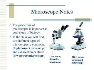
Light Microscopes • Magnifies objects using light • Simple microscopes: have only one lens (magnifying glass) • Compound light microscope: has two lenses
Using the microscope: • Find total magnification: multiply power of eyepiece (usually 10X) x power of objective lens • When focusing: always start with LOW POWER objective • Move nosepiece all the way down and focus slowly UPWARD using the coarse adjustment
Electron Microscopes • 1940’s, uses electron beams instead of light • Allows more detail, but specimens are usually killed in the staining process • Two types: • TEM: beam goes through specimen • SEM: scans surface only
Parts of a microscope • Ocular lens: lens near eye • Eyepiece: holds ocular lens • Objective lens: lens near specimen • a. high power lens: longer, usually 40 X • b. low power lens: shorter, usually 10 X • Nosepiece: holds objective lenses • Stage: platform supporting specimen • Diaphragm: regulates amount of light • Fine adjustment/coarse adjustment: used to focus on object • Arm: supports microscope • Illuminator: light source
How did the Cell Theory come about? • Robert Hooke discovered cells in 1665 • Examined a wine cork(tree) • looked like his monastery cell so he named them “cells”
Schleiden and Schwann (1838-1839) examined many types of cells • Schleiden: botanist, discovered that all plant parts are made of cells • Schwann: zoologist, discovered that all animals parts are made up of cells • Both observed / different types of cells performed different functions Schwann Schleiden
Rudolph Virchow (1855) • Physician that stated all living cells come only from other living cells
The Cell Theory • All organisms are made up of cells • Cell is the basic unit of organization • Cells come from pre-existing cells
Stem Cell Research Website-stem cell animation http://www.sumanasinc.com/webcontent/anisamples/stemcells.html