Download
1 / 31
310 likes | 518 Views
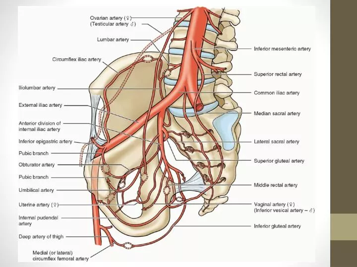
Question. Blood supply of the rectum?. Superior rectal artery (inferior mesenteric) Middle rectal artery (anterior division of internal iliac) Inferior rectal artery (internal pudendal ). Nerves of the Pelvis. Pudendal S2‑S4

E N D
Question • Blood supply of the rectum? • Superior rectal artery (inferior mesenteric) • Middle rectal artery (anterior division of internal iliac) • Inferior rectal artery (internal pudendal)
Nerves of the Pelvis • Pudendal • S2‑S4 • Sensory to genitalia; muscular branches to perineal muscles, external urethral sphincter, external anal sphincter • Pelvic splanchnic • S2‑S4 • Pelvic viscera via inferior hypogastric and pelvic plexuses • Nerve to levator ani and coccygeus • S3‑S4 • Levator ani and coccygeus muscles
Spermatic Cord • Contains structures to and from testes • Begins at deep inguinal ring, lateral to inferior epigastrics • Fascial coverings: • Internal spermatic fascia (transversalisfascia) • Cremasteric fascia (internal oblique) – contains cremaster muscle • External spermatic fascia (external oblique) • Contents: • Ductus (vas) deferens • Testicular artery, cremasteric artery, deferential artery, pampiniform plexus • Sympathetics, genital branch of genitofemoral nerve • Vestige of processus vaginalis • Lymphatics
Anatomy of the Scrotum • Cutaneous sac • Dartos fascia (continuous with Scarpa and Colles fascia) • Dartos muscle • Septum, scrotal raphe • Innervation: Genital branch of genitofemoral nerve (anterolateral surface) • Posterior scrotal nerves (internal pudendal, S2-S4)
Anatomy of Testes • Tunica vaginalis = closed peritoneal sac = closed-off remnant of processus vaginalis in embryo • Testis covered by visceral layer, except for attachment to epididymis and spermatic cord • Parietal layer is adjacent to internal spermatic fascia • Small amount of fluid allows free movement • Tunica albuginea = tough fibrous outer surface • Mediastinum of testis = origin of fibrous septa • Right testicular vein IVC, left vein renal artery
Epididymis • Duct of epididymis = Convoluted tube of smooth muscle of posterior aspect of testes – moves spermatozoa distally with peristalsis and stereocilia • Head superior expanded part (lobules of 12-14 ductuli efferentes) • Appendices of epididymis = remnant of mesonephric (Wolffian) duct • Body • Tail
Ductus Deferens • Continuation of duct of epididymis (from tail) – ascends posterior to testis and medial to epididymis • Penetrates abdominal wall via spermatic cord / inguinal canal, crossing external iliac vessels • Crosses ureter, terminates as the ampulla of ductus deferens to join duct of seminal gland to form ejaculatory duct • Artery to ductus deferens arises from superior vesical
Seminal Vesicles and Ejaculatory Duct • Seminal vesicle (gland) about 5cm-long glandular diverticulum between fundus of bladder and rectum • Does not store sperm • Duct of seminal vesicles joins ampulla of ductus deferens • Ejaculatory duct: 2.5cm long, slender tubes that pass through prostate • Alongside prostatic utricle • Open on the verumontanum (seminal colliculus) • Supplied by artery to ductus deferens • Ductus deferens, seminal glands, ejaculatory ducts and prostate innervated by sympathetic nervous system (intermediolateral cell column T12-L2 via transverse lumbar splanchnic nerves, hypogastric and pelvic nerve plexuses)
Prostate • Fibrous capsule + visceral layer of pelvic fascia = prostatic sheath • Connects with puboprostatic ligaments anteriorly • Rectovesical septum posterior • Base (neck of bladder) • Apex (urethral sphincter, perineal muscles) • Anterior surface (rhabdosphincter/ external urethral sphincter); Space of Retzius / retropubic space • Posterior surface (ampulla of rectum) • Inferolateral surface (levator ani) • Largest accessory gland of male system – 3 x 4 x 2cm (length/width/depth)
Divisions of prostate • Lobes • Isthmus (anterior lobe) • Right and left lateral lobes • Inferoposterior lobule (palpable in DRE) • Inferolateral lobule (lateral to urethra) • Median lobe – undergoes hypertrophy • Superomedial lobule (surrounds ejaculatory duct • Anteromedial lobule (proximal prostatic urethra) • Zones • Transition zone (5% of glandular tissue, around proximal urethra – site of benign nodular hyperplasia) • Central zone (around ejaculatory ducts, 20% of tissue) • Peripheral zone (70% of tissue, most cases of prostatic carcinoma)
Prostatic ducts (20-30) open into prostatic sinuses either side of verumontanum (seminal colliculus) • Prostatic utricle = remnant of uterovaginal canal • Corpora amylacea = inspissated prostatic secretions that increase with age and become calcified • Prostatic arteries = branches of inferior vesicle (some from internal pudendal and middle rectal) • Prostatic venous plexus drains into internal iliac
Male Urethra • Four parts: intramural, prostatic, membranous, spongy • Membranous • Apex of prostate to bulb of penis via external urethral sphincter • Bulbourethral glands of Cowper lie posterolateral to membranous urethra, inside external urethral sphincter • Spongy urethra in corpus spongiosum, 5mm in diameter • Intrabulbar fossa in bulb of penis • Navicular fossa in glans • Urethral glans secrete mucus • Somatic innervation visa dorsal nerve of penis (pudendal) • Dorsal artery of penis supplies membranous and spongy parts
Penis • Erect in anatomical position, so dorsum faces anteriorly when flaccid • Three cylindrical cavernous bodies • Paired corpora cavernosa dorsally (singular = corpus cavernosum). Forma crura of the penis proximally, separated by septum penis • Corpus spongiosum • Each covered in fibrous capsule, tunica albuginea • Root of penis = attached part. Located in superficial perineal pouch • Crura = masses of erectile tissue. Each crus attached to ischial ramus • Bulb = enlarged posterior part. Penetrated by urethra • Ischiocavernosus and bulbospongiosus muscles • Suspensory ligament of penis (sling from symphysis) • Fundiform ligament of penis (from linea alba, anterior to symphysis) • Buck’s fascia = deep fascia of the penis (continuation of perineal fascia)
Body is free part suspended from pubic symphysis. No muscular tissue • Glans = distal expansion of corpus spongiosum • Corona of glans = proximal margin • Coronal sulcus = oblique groove overhung by corona • Covered by prepuce (foreskin) • Frenulum of prepuce = median fold • External urethral meatus near tip of glans • Arterial supply: From internal pudendal • Dorsal arteries of penis run in dorsal groove between corpora cavernosa • Deep arteries run in centre of corpora cavernosa • Form helicine arteries of penis in erectile tissue • Venous drainage: Dorsal vein of penis drains to prostatic venous plexus • Innervation: PSNS S2-S4 via pelvic splanchnic and pudendal