Download
1 / 15
150 likes | 384 Views
How do we classify something as living?. Living things… 1. Have a need for energy (from chemicals, sunlight, other animals, etc.) 2. Respond to their environment (to survive) 3. Have the ability to reproduce= pass genetic info (asexual or sexual) 4. Are made up of one or more cells.

E N D
How do we classify something as living? Living things… 1. Have a need for energy (from chemicals, sunlight, other animals, etc.) 2. Respond to their environment (to survive) 3. Have the ability to reproduce= pass genetic info (asexual or sexual) 4. Are made up of one or more cells
Cell Theory • 3 basic components: • All organisms are made of cell(s). • All existing cells are produced by other living cells. • The cell is the most basic unit of life.
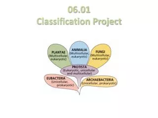
2 Types of Cells • 1. Prokaryotes= No nucleus (“before” nucleus) • Bacteria (Kingdom Monera)
(Kingdom Fungi) • 2. Eukaryotes= Nucleus (“true” nucleus) All other Kingdoms: • Fungi • Plants • Protists • *Animals Eukaryotic cells have many organelles- Structures that perform specific functions. *plasma membrane, cytoplasm, nucleus (Kingdom Plantae) (Kingdom Protista) (Kingdom Animalia)
Cell Locomotion • Cilia = “eyelashes” • Flagella = “small whips” *Locomotion needed to find a food source or escape predators/harsh conditions.
Tools for Viewing Life • Light Microscope • Compound • Stereo/ Dissecting • Electron Microscope • Scanning (SEM) • Transmission (TEM) • X-rays • Ultrasound • MRI = Magnetic resonance imaging
Light Microscopes Compound= 1 optical system *Magnification = Up to 1500x (LHS=400x) (objective x eyepiece) *Images = Upside down and reversed http://ettc.lrhsd.org/archives/Pictures/138-microscopes-lg.jpg http://www.az-microscope.on.ca/images/Ml2100.jpg
Stereo = 2 separate optical systems (for objects that will not fit on a slide) *Magnification = Up to 100x (LHS = 40x) *Images = 3D, normal upright, right to left image http://www.microscopyu.com/articles/stereomicroscopy/stereointro.html http://www.clt.astate.edu/mhuss/stereoparts.jpg
Electron Microscopes • Scanning (SEM) *Uses electrons instead of light to form/focus image *Used to view surfaces of objects (electrons deflect off specimens) *Magnification up to 500,000x Fly Head http://image53.webshots.com/53/8/76/41/2484876410085329142tuvzFM_fs.jpg http://gsc.nrcan.gc.ca/labs/ebeam/images/sem8.jpg
Transmission (TEM) *Uses electrons instead of light to form/focus image *Used to view inner structure of objects (electrons pass through specimens) *Magnification up to 1, 000,000x TEM-micrograph: thylakoid system in a chloroplast (bar= 0.5 µm). http://www.iopb.res.in/~bhupen/tem_mch.gif http://images.google.com/imgres?imgurl=http://www-classic.uni-graz.at/pphwww/elmi/tem2.jpg&imgrefurl=http://www-classic.uni-graz.at/pphwww/elmi/tempraeparatione.htm&usg=__a3m7XGYsX7CRKvKD0cd0qlD36jk=&h=321&w=387&sz=43&hl=en&start=21&um=1&tbnid=Ugh374nux3hqcM:&tbnh=102&tbnw=123&prev=/images%3Fq%3DTEM%26ndsp%3D18%26hl%3Den%26sa%3DN%26start%3D18%26um%3D1
TEM vs. SEM http://www.vcbio.science.ru.nl/images/TEM-SEM-electron-beam.jpg
X-Ray • X-rays pass through tissue to show dense material (which absorbs the rays) * CT- scans also use this technique http://www.designswan.com/wp-content/uploads/2008/xray/22.jpg http://www.antonine-education.co.uk/physics_gcse/Unit_1/Topic_5/em_spectrum.jpg
Ultrasound • High-frequency sound waves pass through the body until they come to a border between two tissues that conduct sound differently. Then, some of the sound waves bounce back & are produced as a picture. *When used for long periods of time at high intensities, it can cause the tissues to become heated. http://www.nlm.nih.gov/medlineplus/ency/images/ency/fullsize/18056.jpg http://www.hip2b2.com/images/uploaded_images/Ultrasound.jpg
MRI • Uses a magnetic field and radio waves to create detailed images of body tissues. *Can take images from almost every angle http://i.ehow.com/images/GlobalPhoto/Articles/4673792/92555-main_Full.jpg http://www.magnet.fsu.edu/education/tutorials/magnetacademy/mri/images/mri-scanner.jpg