PROTEIN
PROTEIN. Elements: C, H, O, N , ( S, P). Kuliah ke-4 BIOKIMIA FAKULTAS PETERNAKAN UNIVERSITAS BRAWIJAYA Prof. Dr.sc.agr. Ir. Suyadi, MS. INTRODUCTION: PROTEIN. Elements: C, H, O, N, and sometimes S.

PROTEIN
E N D
Presentation Transcript
PROTEIN Elements:C, H, O, N, (S, P) Kuliah ke-4 BIOKIMIA FAKULTAS PETERNAKAN UNIVERSITAS BRAWIJAYA Prof. Dr.sc.agr. Ir. Suyadi, MS.
INTRODUCTION: PROTEIN • Elements: C, H, O, N, and sometimes S. • Function: Enzymes, structural proteins, storage proteins, transport proteins, hormones, proteins for movement, protection, and toxins.
Introduction • The subunits of a protein are amino acids or to be precise amino acid residues. • An amino acid consists of: • a central carbon atom (the alpha Carbon Calpha) and an amino group (NH2), • a hydrogen atom (H), • a carboxy group (COOH) and • a side chain (R) which are bound to the Calpha.
General Structure • Proteins are made from several amino acids, bonded together. It is the arrangement of the amino acid that forms the primary structure of proteins. The basic amino acid form has a carboxyl group on one end, a methyl group that only has one hydrogen in the middle, and a amino group on the other end. Attached to the methyl group is a Rgroup.
General Structure • Proteins are made from several amino acids, bonded together. It is the arrangement of the amino acid that forms the primary structure of proteins. • The basic amino acid form has a carboxyl group on one end, a methyl group that only has one hydrogen in the middle, and a amino group on the other end. • Attached to the methyl group is a Rgroup.
General Structure There are 20+ amino acids, each differing only in the composition of the R groups. An R group could be a sulfydrl, another methyl, a string a methyls, rings of carbons, and several other organic groups. Proteins can be either acidic or basic, hydrophilic or hydrophobic. The following table shows 20 amino acids that common in proteins.
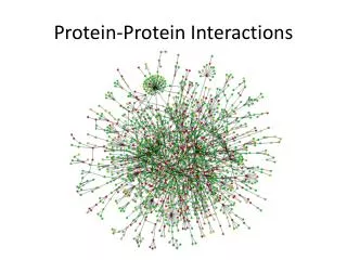
Proteins play key roles in a living system • Three examples of protein functions • Catalysis:Almost all chemical reactions in a living cell are catalyzed by protein enzymes. • Transport:Some proteins transports various substances, such as oxygen, ions, and so on. • Information transfer:For example, hormones. Alcohol dehydrogenase oxidizes alcohols to aldehydes or ketones Haemoglobin carries oxygen Insulin controls the amount of sugar in the blood
R NH3+ C COO- H Amino acid: Basic unit of protein Different side chains, R, determin the properties of 20 amino acids. Amino group Carboxylic acid group An amino acid
20 Amino acids Leucine (L) Isoleucine (I) Valine (V) Alanine (A) Glycine (G) Proline (P) Asparagine (N) Methionine (M) Tryptophan (W) Phenylalanine (F) Tyrosine (Y) Threonine (T) Serine (S) Cysteine (C) Glutamine (Q) Histidine (H) Glutamic acid (E) Arginine (R) Asparatic acid (D) Lysine (K) White: Hydrophobic,Green: Hydrophilic,Red: Acidic,Blue: Basic
Proteins are linear polymers of amino acids R1 R2 COOー COOー NH3+ NH3+ + + C C H H A carboxylic acid condenses with an amino group with the release of a water H2O H2O R1 R2 R3 C C CO CO C CO NH3+ NH NH Peptide bond Peptide bond H H H The amino acid sequence is called as primary structure D F T A A S K G N S G
・ G C G C T T A A G C G C ・ ・ C G C G A A T T C G C G ・ Amino acid sequence is encoded by DNA base sequence in a gene DNA molecule DNA base sequence =
Amino acid sequence is encoded by DNA base sequence in a gene
Gene Gene Gene Gene Gene Gene Gene Gene Gene Gene Gene Gene Gene Gene Protein Protein Protein Protein Protein Protein Protein Protein Protein Protein Protein Protein Protein Protein Gene is protein’s blueprint, genome is life’s blueprint DNA Genome Gene Protein
Gene Gene Gene Gene Gene Gene Gene Gene Gene Gene Gene Gene Gene Gene Protein Protein Protein Protein Protein Protein Protein Protein Protein Protein Protein Protein Protein Protein Gene is protein’s blueprint, genome is life’s blueprint Glycolysis network Genome
In 2003, Human genome sequence was deciphered! • Genome is the complete set of genes of a living thing. • In 2003, the human genome sequencing was completed. • The human genome contains about 3 billion base pairs. • The number of genes is estimated to be between 20,000 to 25,000. • The difference between the genome of human and that of chimpanzee is only 1.23%! 3 billion base pair => 6 G letters & 1 letter => 1 byte The whole genome can be recorded in just 10 CD-ROMs!
Each Protein has a unique structure Amino acid sequence NLKTEWPELVGKSVEEAKKVILQDKPEAQIIVLPVGTIVTMEYRIDRVRLFVDKLDNIAEVPRVG Folding!
Basic structural units of proteins: Secondary structure α-helix β-sheet Secondary structures, α-helix and β-sheet, have regular hydrogen-bonding patterns.
Three-dimensional structure of proteins Tertiary structure Quaternary structure
Hierarchical nature of protein structure Primary structure (Amino acid sequence) ↓ Secondary structure (α-helix, β-sheet) ↓ Tertiary structure (Three-dimensional structure formed by assembly of secondary structures) ↓ Quaternary structure (Structure formed by more than one polypeptide chains)
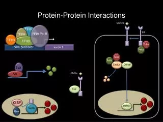
Close relationship between protein structure and its function Antibody Hormone receptor Example of enzyme reaction substrates A enzyme enzyme B Matching the shape to A Digestion of A! enzyme A Binding to A
Protein structure prediction has remained elusive over half a century “Can we predict a protein structure from its amino acid sequence?” Now, impossible!
Protein Classification • Proteins can be described as having several layers of structure. At the lowest level, the primary structure of proteins are nothing more that the amino acids which compose the protein, and how those proteins are bonded to each other. The bonds between proteins are called peptide bonds, and they can have either single bonds, double bonds, triple bonds, or more holding the amino acids into a protein molecule. • At the next level, the secondary structure of proteins, proteins show a definite geometric pattern. One pattern that the protein can take is a helical structure, similar to a spiral staircase. Hair has such a secondary structure. When examined closely, you can see the turns in the proteins of hair molecules. A second geometric pattern is the pleated sheet, where several polypeptide chains go in several different directions. I think of a sheet of paper, or a length of fabric. When viewed closely, silk fibronin, the silk protein, forms such a shape. Skin, although made of more than just proteins, provides another example of a protein with a sheet structure. The following figure shows the pleated sheet secondary structure of silk.
Contoh struktur protein • Skin fibroin
Next, we find a tertiary structure to proteins. Here, we find the three-dimensional structure of the globular proteins, where disulfide bridges puts kinks and bends in the secondary structure. Again thinking about hair, some people have straight hair, some have wavy hair, and some have curly hair. The links and bends in the secondary structure causes the curls in hair. Curly hair has more kinks and bends that wavy hair, and straight hair has very few, if any bends
Contoh struktur protein • Psoriasin
At the last, we see the quaternary structure of proteins. This the the form taken by complex proteins formed from two or more smaller, polypeptide chains. The polypeptide chains form pieces of a jigsaw puzzle, that when put together form a single protein. Hemoglobin provides a good example, being made from four polypeptide chains.
Contoh struktur protein • Hemoglobin
Protein Function in Cell • Enzymes • Catalyze biological reactions • Structural role • Cell wall • Cell membrane • Cytoplasm
Hemoglobin: Background • Protein in red blood cells
Hemoglobin: Background • Protein in red blood cells • Composed of four subunits, each containing a heme group: a ring-like structure with a central iron atom that binds oxygen Red blood cell
Hemoglobin: Background • Protein in red blood cells • Composed of four subunits, each containing a heme group: a ring-like structure with a central iron atom that binds oxygen • Picks up oxygen in lungs, releases it in peripheral tissues (e.g. muscles)
Hemoglobin – Quaternary Structure Two alpha subunits and two beta subunits (141 AA per alpha, 146 AA per beta)
Hemoglobin – Tertiary Structure One beta subunit (8 alpha helices)
Hemoglobin – Secondary Structure alpha helix
Structure Stabilizing Interactions • Noncovalent • Van der Waals forces (transient, weak electrical attraction of one atom for another) • Hydrophobic (clustering of nonpolar groups) • Hydrogen bonding
Hydrogen Bonding • Involves three atoms: • Donor electronegative atom (D) (Nitrogen or Oxygen in proteins) • Hydrogen bound to donor (H) • Acceptor electronegative atom (A) in close proximity D – H A
δ- δ+ δ- D – H A D-H Interaction • Polarization due to electron withdrawal from the hydrogen to D giving D partial negative charge and the H a partial positive charge • Proximity of the Acceptor A causes further charge separation
δ- δ+ δ- D – H A D-H Interaction • Polarization due to electron withdrawal from the hydrogen to D giving D partial negative charge and the H a partial positive charge • Proximity of the Acceptor A causes further charge separation • Result: • Closer approach of A to H • Higher interaction energy than a simple van der Waals interaction
Hydrogen Bonding And Secondary Structure beta-sheet alpha-helix