Download

1 / 48
500 likes | 739 Views
This article explores how animals acquire their food through autotrophic and heterotrophic mechanisms. Autotrophs create energy-rich molecules using sunlight or inorganic chemicals, while heterotrophs depend on autotrophs for sustenance. The piece details various feeding mechanisms such as filter feeding, substrate feeding, fluid feeding, and bulk feeding. It also provides an overview of the human digestive system, including the roles of major organs like the mouth, esophagus, stomach, and intestines in the processes of ingestion, digestion, absorption, and elimination.

E N D
Digestive System II SBI3U Ms. De Sousa
How AnimalsObtaintheirfood Autotrophs: • Take energy from the environment in the form of sunlight or inorganicchemicals • Use the energy to create energy-rich molecules. • Examples: plants and algae
How AnimalsObtaintheir Food Heterotrophs: • Take in autotrophsas food. • Depend on autotrophs for the energy and raw materials they need • Heterotrophs obtain energy by breaking down organic molecules obtained from the autotrophs • Examples: all animals, mostfungi, bacteria and protozoa.
Animals (i.e. heterotophs) • Have adaptedmechanisms to search, obtain and take in theirfood. • Animalsobtainfoodfromeither of the following 4 mechanisms.
FeedingMechanisms 1) FilterFeeding: • Use a filter basket to obtainorganismssuspended in the water. • Siphon water intoitsmouth and filtersit to obtainitsfood. Examples: Tube worms, clams, whales
FilterFeeding in Sponges Video
FeedingMechanisms 2) SubstrateFeeding: • Live within or on the food source and eattheirwaythroughit. • Examples : Earthworms, caterpillars
FeedingMechanisms 4) Bulk Feeders: • Most vertebrates (includinghumans). • Ingest large pieces of food or swallowitwhole. • Differentanimalsmay use tentacles, pincers and claws to eat.
FeedingMechanisms 3) Fluid Feeders: • Suck or lickfluidsform plants or animals. • Theirmouth parts are adapted to piercethrough skin or leaf tissue. • Examples: spidders, bees, butterflies.
Digestive Tract • In order for the cells to obtainnutrients, the digestive system needs to break them down intosmall, soluble units. • The smallunits of nutrientsdiffuse intocell membranes and into the circulatory system.
Alimentary Canal • Most animals have a digestive tract alsoknown as « Alimentary Canal » • Alimentary Canal: tube wherefood in processed, beginningat the mouth and endingat the anus. • There are variousorgansalong the alimentary canal thatprocess the food.
Alimentary Canal Pouchlike structure thatsoftens and stores food Example: Earthworm Churns and grinds the food
All organs in the digestive system play a vital role in the process of digestion. Eachorganisresponsible for eitherbreaking down the food, abosrbingit or deliveringit to other areas.
Similarities and Differences • The function of the alimentary canal is the same for all animals. • The lengthdiffersaccording to feeding habits. • Herbivores and Omnivores have longer digestive tracts thanCarnivores.
Stages of Digestion • No matter the feedingmechanisms all animalsundergoe the same 4 stages of digestion. a) Ingestion b) Digestion c) Absorption d) Elimination
Stages of Digestion • Ingestion:Taking in the food • Digestion: Breakdown of foodintosmallmolecules • Absorption: diffusion of smallmoleculesinto the circulatory system • Elimination: removal of undigestedsolidwastefrom the body.
Main Types of Digestion: Mechanical: physical breakdown of macronutrients (i.e. chewing, churning) Chemical: chemical breakdown of nutrientsintosmallermolecules by hydrolysisand enzymatic action.
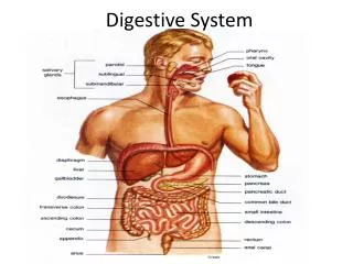
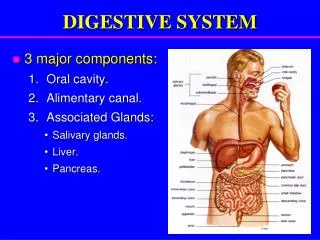
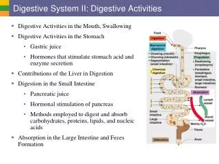
10.2 The Human Digestive System The digestive tract has numerousorganswithspecificfunctions. Eachorganhelpsto breakdown food.
DIAGRAM OF DIGESTIVE SYSTEM: Mouth Esophagus Liver Stomach Pancreas Gallbladder Small Intestine Large Intestine Rectum Appendix Anus
STEP 1: INGESTION Mouth Mechanical digestion: Teethbreakdown the foodintosmallpieces Chemical digestion: Amylase (enzyme) breaks down the bonds in carbohydrates.
Salivaissecretedfrom 3 salivaryglands. • The secretion of saliva istriggeredbeforeyou have food in yourmouth.
Role of Saliva (Chemical Digestion) • Amylase (enzyme) breaks down carbohydratesintosimplersugars. • Dissolves water soluble foodparticles • Stimulates taste buds. • Lubricates the foodsoitcanbeswallowed.
When the mouth has created a bolus of food, the tongue pushes it back into the back of the throat. • Epiglottis – flap covers trachea so food doesn’t get in • Food stretches walls of esophagus and moves downward through waves of contractions called peristalsis
Esophagus • Glands in the liningproducemucuswhichkeeps the tube moist and facilitatesmovement of food. • The muscles in the esophaguscontractinvoluntarily to push the bolus of foodinto the stomach. Video
Esophagus • When the bolus of food has reached the bottom of the esophagus the EsophagealSphincter opens. • Whileswallowing, the muscles relax, opening the sphincter and allowing the food to enter the stomach.
The esophageal sphincter usuallyremainsclosed to preventacidicjuicesfromflowing up into the esophagus. Acid Reflux Video
STEP 2: DIGESTION Stomach • Muscular, J-shapedorgan • Leftside of the abdominal cavitybelow the diaphragm • Performsbothchemical and mechanical digestion
Stomach • The walls of the stomach are foldedsothatitcanexpandafter a meal. • The glands on the stomachwall release « gastricjuice » whichconsist of HCl, salts, enzymes, water and mucus. (chemical digestion) • The walliscovered in a mucus coatwhichprotectsitfrom the acidreleasedfrom the gastricjuices.
Stomach Pepsinis the enzyme released. Pepsinremains inactive (pepsinogen) untilHClissecretedform the glands. Once activated, pepsinhydrolyzesproteins.
Stomach • The HCl breaks down food and destroys foreignbacteria in the food. • Stomachcontracts and relaxes to churn the food. • The churninghelpsto break up the foodand mix the gastricjuices. • Food + Gastricjuices = Chyme
Pyloric Sphincter The Pyloric Sphincter opens to move the chyme into the small intestine.
STEP 2: DIGESTION Cont’d Small Intestine • Furtherbreaks down and abosrbsnutrients. • Composed of three main components: 1) Duodenum 2) Jejunum 3) Illeum
Duoedenum • Receivessecretions(enzymes) fromthe gallbladder and pancreasto further breakdown nutrients. • The intestinal glands also release trypsin and chymotrypsin (enzymes) to breakdown carbohydrates and proteins. • Breaks down proteins, fats and carbohydrates.
Jejunum • 2.5 m long. • Contains more foldsthan the duodenum. • Breaksdown the remainingproteins and carbohydratessothatitcanbeabsorbed.
Villi and Microvilli • Small intestine iscoveredby tinyfinger-likeprojections called« Villi » • The villiincreases the surface area for absorption of nutrientsinto the bloodstream. • There is a capillary network within the villi. The nutrients diffuse from the small intestine into the villiwherebyitthen diffuses into the capillary network.
Illeum • 3 m long. • Has fewervillithan the dueodenum and jejunum. • Absorbsnutrientsthrough the villiand pushesundigestedmaterialinto the large intestine.
STEP 3: ABSORPTION Small Intestine and Large intestine The villi and microvilliincrease surface area for absorption. The nutrients diffuse into the capillarieswithin the villi.
STEP 3: ABSORPTION Small Intestine and Large intestine • Large intestine (colon) is 1.5 m in length • Reasborbs90% of water and electrolytesback into the blood. • Bacterialive hereproducingvitamin K /B and break down undigestedmatter. • Anyundigestedmaterialthatremainsiscalledfeces. • Fecalmatterisstoredhere for eliminationthrough the rectum.
STEP 4: ELIMINATION Rectum Main component of feces: • Cellulose- makes up plant cell walls, cannot be digested by humans • Living and dead bacteria • Water • Toxic wastes are removed • People who don’t eat enough cellulose (plant material and fibre) have fewer bowel movements and are at risk of colon cancer.
Pancreas • Secretes about 1 L of pancreaticfluidinto the duoedenumeachday. • PancreaticFluid: 1) Lipase (enzyme) – chemically digest lipids 2) Bicarbonate– alterspH of chyme sothatenzymescanbeactivated. (pH 1 to pH 8)
Liver • Largestinternalorganin the human body. • Releases bile (greenish-yellowfluid made up of bile pigments and salts) • Bile is sent to the gallbladderwhereitistemporarilystored.
Bile • Fats are insoluble in water and remainsuspended in the chyme. • The bile salts break up the fat dropletssotheycan disperse through the chyme. • The enzymes break the dropletsapart. Video http://www.youtube.com/watch?v=Z7xKYNz9AS0
Enzymes – proteinsthat speed up chemicalreactions Digestive Enzymes Induced Fit Model The substrate and enzyme have complementaryshapes. Thusmakingthem fit perfectlyinto one another. Therefore, the enzyme ishighlyspecific to itssubstrate.