Comprehensive Guide to Body Anatomy and Microscopy Techniques
180 likes | 255 Views
Explore external/internal body structures, body cavities, and imaging methods in anatomy. Understand cells, tissues, organs, systems, and microscopes' workings like light and electron microscopy.

Comprehensive Guide to Body Anatomy and Microscopy Techniques
E N D
Presentation Transcript
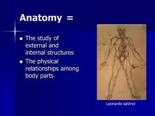
Anatomy = • The study of external and internal structures • The physical relationships among body parts Leonardo daVinci
Atoms Molecules and Macromolecules, such as proteins Organelles Cells Tissues Organs Organ Systems Organism Organization
Planes • Frontal • Think front! • Transverse • “ Trans” = across • Sagittal • Especially mid-sagittal
Quadrants Surrounding the Umbilicus • RUQ: Gall Bladder, Rt kidney • RLQ: Appendix, Rt ovary • LUQ: Lt liver lobe, stomach, Lt kidney • LLQ: Small intestine, Lt ovary • Superficial: Toward the skin • Deep: Farther inside
These words are used all the time, including this class! Note the Anatomic Position.
Body Cavity Membranes More later!
Imaging • Light Microscopy (cytology) • Electron Microscopy (ultrastructure) • TEM • SEM • Radiography (X-Rays) • CT Scanning • Ultrasound • Magnetic Resonance Imaging (MRI)
Light Microscopy • The specimen is fixed • Usually in formalin • Then embedded in paraffin • Sectioned with a microtome • Approx 5 μ sections • and stained • Enhances contrast for better visualization • Many types of stains • H & E = Hematoxylin and Eosin • May add “artifact”
Concepts Important for Viewing Resolution -The distance between two objects that is required for the two objects to be distinguished. Depth of Field -depth that focus is clear Contrast Formation -(e.g. absorption contrast) Illumination Source -diascopic vs. episcopic from below (compound) vs. from above (dissecting) Artifact – Distortion from preparation of the specimen
How The Concepts Interact As Resolution and Brightness improve, Depth of Field and Contrastare diminished. Vice versa is also true. These are controlled by the iris diaphragm, the position of the condensor, and the magnification.
Electron Microscopy Transmission Electron Microscope Uses a beam of electrons to view topography, morphology, composition, and crystallography. EM was developed for 10,000 X magnification. Properties of light limit magnification of light microscopes to 1000 X and resolution to 0.2 m.