Download
1 / 1
10 likes | 159 Views
A Biomimetic Electrostatic Imaging System Proposed for Real-time In-vivo Surgical Guidance of Tumor Resection Jonathan Friedman, PhD, Peyman Golshani, MD/PhD, Mani Srivastava, PhD

E N D
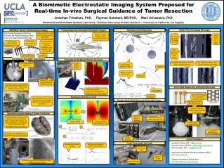
A Biomimetic Electrostatic Imaging System Proposed for Real-time In-vivo Surgical Guidance of Tumor Resection Jonathan Friedman, PhD, Peyman Golshani, MD/PhD, Mani Srivastava, PhD Networked and Embedded Systems Laboratory / Golshani Laboratory for Neuroscience --- University of California, Los Angeles . Golshani Lab Department of Neurology David Geffen School of Medicine, UCLA Calibration and Compensation World’s First 16 Electrode BEI Images ABSTRACT: Electric Field Disturbance Imaging Proposed Approach: Biomimetic Electrostatic Imaging Goal: Provide Simultaneous Real-Time Functional and Anatomical Imaging of Brain Tissue Using Quasi-Static Electric Fields Conductivity of Tumor Tissue is 10x Lower than Normal Brain Tissue “Electric” Fish Detect Changes to a Self-Generated Electric Field White Pixels Show Areas of Increased Conductivity Cross Section View of Tank at Depth Determine Spatial Conductivity of Tissue from Disturbance to Self-Generated Electric Field (like MRI) Surface Probe (non-destructive) State of the Art Neuroprobes Destroy Tissue and Cannot Image Actual Location of Pipe Target (Yellow Circle) The Brain! Current Proposed The Problem: “Inoperable” Brain Tumors Generating the Background Field (in the Ocean) Pipe Target Nearing Edge of Electrode Array – Accuracy Degrades Pipe Target Beyond Sensor Array – Accuracy Degrades Substantially Tumor Clearly Visible on MRI, BUT… Custom Gantry & Cantilever Induced Dipoles Rotate in the Field Expected Model is Fit to Tank Measurements Observed …MRI Structure and RF Transducers Preclude Real-time Use in OR Stereotactic Camera Systems Register the Patient to the MRI – ineffective once the tissue resection begins Future Work: Reduce Size and Scale of Electrodes • >10,000 Sample Points • Over 6 Hours of Measurement • Highest Resolution Study Published Compensation Calibration High Density AlphaSTAR Coated Electrodes Modeling the Disturbance Field Better Boundary Guidance Would Yield Better Outcomes & Enable Resections Currently Considered Too High Risk Results: World’s First Biomimetic Electrostatic Imager Inexpensive Disposable Flex-PCB Electrodes Preparing for Real-time Imaging in Awake Behaving Animal Models Mark M. Souweidane, MD NewYork-Presbyterian Hospital/Weill Cornell Medical Center Real-time Functional Imaging of Cardiac Conduction Each Card Supports 17 Electrodes The Same Approach Could Be Used to Visualize the Conductive (Purkinje) Fibers -- Reducing 8 Hour Guess-and-Check Catheterization Lab Procedures to Less Than One Hour While Guaranteeing Efficacy of Treatment Multiple BEI Units Can Connect to the Same Host Conductive Object (Induced Dipole) In Field Background Field 2-Photon uScope Provides Reference 1 2 Craniotomy for Electrode Access Fluroscope Uses Ionizing Radiation – Proposed Approach is Intrinsically Safe Rig Images Courtesy Jiyoung Park 3 68 Total Electrodes Contact Information Jonathan Friedman, PhD – jf@ee.ucla.edu Peyman Golshani, MD/PhD – pgolshani@mednet.ucla.edu Mani Srivastava, PhD – mbs@ee.ucla.edu Networked and Embedded Systems Laboratory (NESL) http://nesl.ee.ucla.edu Golshani Laboratory for Neuroscience http://golshanilab.neurology.ucla.edu/ “Large” Scale Target Tank Trials Disturbance Field Caused by Object Disturbance as Observed by Different Electrode Pairs Gantry Moves Target Metal Pipe (Target) Fluroscope Imagery is Non-Functional and Cannot Visualize the Relevant Anatomy Imager’s Electrodes