Download
1 / 130
1.3k likes | 1.65k Views
Explore how the immune system's innate and adaptive defenses work to protect the body from pathogens. Learn about phagocytes, natural killer cells, inflammation, and more. Discover the importance of surface barriers like skin and mucous membranes in warding off invaders.

E N D
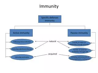
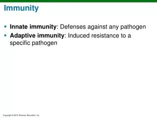
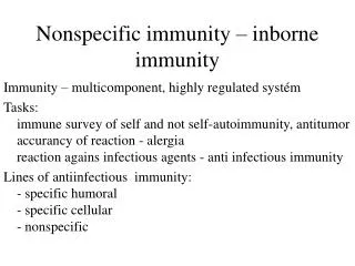
Immunity • Resistance to disease • Immune system • Two intrinsic systems • Innate (nonspecific) defense system • Adaptive (specific) defense system
Immune System • Functional system rather than organ system • Innate and adaptive defenses intertwined • Release and recognize many of same defensive molecules • Innate defenses do have specific pathways for certain substances • Innate responses release proteins that alert cells of adaptive system to foreign molecules
Immunity • Innate defense system has two lines of defense • First - external body membranes (skin and mucosae) • Second - antimicrobial proteins, phagocytes, and other cells • Inhibit spread of invaders • Inflammation most important mechanism
Immunity • Adaptive defense system • Third line of defense attacks particular foreign substances • Takes longer to react than innate system
Figure 21.1 Overview of innate and adaptive defenses. Surface barriers • Skin • Mucous membranes Innate defenses Internal defenses • Phagocytes • Natural killer cells • Inflammation • Antimicrobial proteins • Fever Humoral immunity • B cells Adaptive defenses Cellular immunity • T cells
Innate Defenses • Surface barriers ward off invading pathogens • Skin, mucous membranes, and their secretions • Physical barrier to most microorganisms • Keratin resistant to weak acids and bases, bacterial enzymes, and toxins • Mucosae provide similar mechanical barriers
Surface Barriers • Protective chemicals inhibit or destroy microorganisms • Acidity of skin and secretions – acid mantle – inhibits growth • Enzymes - lysozyme of saliva, respiratory mucus, and lacrimal fluid – kill many microorganisms • Defensins – antimicrobial peptides – inhibit growth • Other chemicals - lipids in sebum, dermcidin in sweat – toxic
Surface Barriers • Respiratory system modifications • Mucus-coated hairs in nose • Cilia of upper respiratory tract sweep dust- and bacteria-laden mucus toward mouth • Surface barriers breached by nicks or cuts - second line of defense must protect deeper tissues
Internal Defenses: Cells and Chemicals • Necessary if microorganisms invade deeper tissues • Phagocytes • Natural killer (NK) cells • Antimicrobial proteins (interferons and complement proteins) • Fever • Inflammatory response (macrophages, mast cells, WBCs, and inflammatory chemicals)
Phagocytes • Neutrophils most abundant but die fighting • Become phagocytic on exposure to infectious material • Macrophages develop from monocytes – chief phagocytic cells – robust cells • Free macrophages wander through tissue spaces, e.g., alveolar macrophages • Fixed macrophages permanent residents of some organs; e.g., Kupffer cells (liver) and microglia (brain)
Mechanism of Phagocytosis • Phagocyte must adhere to particle • Some microorganisms evade adherence with capsule • Opsonization marks pathogens—coating by complement proteins or antibodies • Cytoplasmic extensions bind to and engulf particle in vesicle called phagosome • Phagosome fuses with lysosome phagolysosome
Figure 21.2a Phagocytosis. Innate defenses Internal defenses A macrophage (purple) uses its cytoplasmic extensions to pull rod-shaped bacteria (green) toward it. Scanning electron micrograph (4800x).
Figure 21.2b Phagocytosis. Slide 1 1 Phagocyte adheres to pathogens or debris. 2 Phagocyte forms pseudopods that eventually engulf the particles, forming a phagosome. Phagosome (phagocytic vesicle) Lysosome 3 Lysosome fuses with the phagocytic vesicle, forming a phagolysosome. Acid hydrolase enzymes 4 Lysosomal enzymes digest the particles, leaving a residual body. 5 Exocytosis of the vesicle removes indigestible and residual material. Events of phagocytosis.
Mechanism of Phagocytosis • Pathogens killed by acidifying and digesting with lysosomal enzymes • Helper T cells cause release of enzymes of respiratory burst, which kill pathogens resistant to lysosomal enzymes by • Releasing cell-killing free radicals • Producing oxidizing chemicals (e.g., H2O2) • Increasing pH and osmolarity of phagolysosome • Defensins (in neutrophils) pierce membrane
Natural Killer (NK) Cells • Nonphagocytic large granular lymphocytes • Attack cells that lack "self" cell-surface receptors • Induce apoptosis in cancer cells and virus-infected cells • Secrete potent chemicals that enhance inflammatory response
Inflammatory Response • Triggered whenever body tissues injured • Prevents spread of damaging agents • Disposes of cell debris and pathogens • Alerts adaptive immune system • Sets the stage for repair
Inflammatory Response • Cardinal signs of acute inflammation: • Redness • Heat • Swelling • Pain (Sometimes 5. Impairment of function)
Inflammatory Response • Begins with chemicals released into ECF by injured tissues, immune cells, blood proteins • Macrophages and epithelial cells of boundary tissues bear Toll-like receptors (TLRs) • 11 types of TLRs recognize specific classes of infecting microbes • Activated TLRs trigger release of cytokines that promote inflammation
Inflammatory Response • Inflammatory mediators • Kinins, prostaglandins (PGs), and complement • Dilate local arterioles (hyperemia) • Causes redness and heat of inflamed region • Make capillaries leaky • Many attract leukocytes to area • Some have inflammatory roles
Inflammatory Response: Edema • Capillary permeability exudate to tissues • Fluid containing clotting factors and antibodies • Causes local swelling (edema) • Swelling pushes on nerve endings pain • Pain also from bacterial toxins, prostaglandins, and kinins • Moves foreign material into lymphatic vessels • Delivers clotting proteins and complement
Inflammatory Response • Clotting factors form fibrin mesh • Scaffold for repair • Isolates injured area so invaders cannot spread
Figure 21.3 Inflammation: flowchart of events. Innate defenses Internal defenses Initial stimulus Physiological response Signs of inflammation Tissue injury Result Release of leukocytosis- inducing factor Release of inflammatory chemicals (histamine, complement, kinins, prostaglandins, etc.) Leukocytosis (increased numbers of white blood cells in bloodstream) Increased capillary permeability Arterioles dilate Attract neutrophils, monocytes, and lymphocytes to area (chemotaxis) Leukocytes migrate to injured area Local hyperemia (increased blood flow to area) Capillaries leak fluid (exudate formation) Margination (leukocytes cling to capillary walls) Diapedesis (leukocytes pass through capillary walls) Leaked clotting proteins form interstitial clots that wall off area to prevent injury to surrounding tissue Leaked protein-rich fluid in tissue spaces Phagocytosis of pathogens and dead tissue cells (by neutrophils, short-term; by macrophages, long-term) Heat Redness Swelling Pain Pus may form Temporary fibrin patch forms scaffolding for repair Locally increased temperature increases metabolic rate of cells Possible temporary impairment of function Area cleared of debris Healing
Phagocyte Mobilization • Neutrophils lead; macrophages follow • As attack continues, monocytes arrive • 12 hours after leaving bloodstream macrophages • These "late-arrivers" replace dying neutrophils and remain for clean up prior to repair • If inflammation due to pathogens, complement activated; adaptive immunity elements arrive
Phagocyte Mobilization • Steps for phagocyte mobilization • Leukocytosis: release of neutrophils from bone marrow in response to leukocytosis-inducing factors from injured cells • Margination: neutrophils cling to walls of capillaries in inflamed area in response to CAMs • Diapedesis of neutrophils • Chemotaxis: inflammatory chemicals (chemotactic agent) promote positive chemotaxis of neutrophils
Figure 21.4 Phagocyte mobilization. Slide 1 Innate defenses Internal defenses Inflammatory chemicals diffusing from the inflamed site act as chemotactic agents. Chemotaxis. Neutrophils follow chemical trail. 4 Capillary wall Basement membrane Endothelium 2 1 3 Leukocytosis. Neutrophils enter blood from bone marrow. Margination. Neutrophils cling to capillary wall. Diapedesis. Neutrophils flatten and squeeze out of capillaries.
Antimicrobial Proteins • Interferons (IFNs) and complement proteins • Attack microorganisms directly • Hinder microorganisms' ability to reproduce
Interferons • Family of immune modulating proteins • Have slightly different physiological effects • Viral-infected cells secrete IFNs (e.g., IFN alpha and beta) to "warn" neighboring cells • IFNs enter neighboring cells produce proteins that block viral reproduction and degrade viral RNA • IFN alpha and beta also activate NK cells
Interferons • IFN gamma (immune interferon) • Secreted by lymphocytes • Widespread immune mobilizing effects • Activates macrophages • Since IFNs activate NK cells and macrophages, indirectly fight cancer • Artificial IFNs used to treat hepatitis C, genital warts, multiple sclerosis, hairy cell leukemia
Figure 21.5 The interferon mechanism against viruses. Slide 1 Innate defenses Internal defenses Virus Viral nucleic acid 1 New viruses Virus enters cell. 5 Antiviral proteins block viral reproduction. Antiviral mRNA 2 Interferon genes switch on. DNA Nucleus mRNA for interferon 4 Interferon binding stimulates cell to turn on genes for antiviral proteins. 3 Cell produces interferon molecules. Interferon receptor Interferon Host cell 2 Host cell 1 Binds interferon from cell 1; interferon induces synthesis of protective proteins Infected by virus; makes interferon; is killed by virus
Complement System (Complement) • ~20 blood proteins that circulate in inactive form • Include C1–C9, factors B, D, and P, and regulatory proteins • Major mechanism for destroying foreign substances • Our cells contain complement activation inhibitors
Complement • Unleashes inflammatory chemicals that amplify all aspects of inflammatory response • Kills bacteria and certain other cell types by cell lysis • Enhances both innate and adaptive defenses
Complement Activation • Three pathways to activation • Classical pathway • Antibodies bind to invading organisms and to complement components • Called complement fixation • First step in activation; more details later
Complement • Lectin pathway • Lectins - produced by innate system to recognize foreign invaders • When bound to foreign invaders can also bind and activate complement • Alternative pathway • Triggered when activated C3, B, D, and P interact on surface of microorganisms
Complement Activation • Each pathway involves activation of proteins in an orderly sequence • Each step catalyzes the next • Each pathway converges on C3, which cleaves into C3a and C3b • Common terminal pathway initiated that • Enhances inflammation, promotes phagocytosis, causes cell lysis
Complement Activation • Cell lysis begins when • C3b binds to target cell insertion of complement proteins called membrane attack complex (MAC) into cell's membrane • MAC forms and stabilizes hole in membrane influx of water lysis of cell • C3b also causes opsonization • C3a and other cleavage products amplify inflammation • Stimulate mast cells and basophils to release histamine • Attract neutrophils and other inflammatory cells
Figure 21.6 Complement activation. Classical pathway Lectin pathway Alternative pathway Activated spontaneously. Lack of inhibitors on microorganism’s surface allows process to proceed Activated by antibodies coating target cell Activated by lectins binding to specific sugars on microorganism’s surface Together with other complement proteins and factors C3 C3a C3b C3b Enhances inflammation: Opsonization: C5b C5a Stimulates histamine release, increases blood vessel permeability, attracts phagocytes by chemotaxis, etc. Coats pathogen surfaces, which enhances phagocytosis C6 MAC C7 C8 C9 MACs form from activated complement components (C5b and C6–C9) that insert into the target cell membrane, creating pores that can lyse the target cell. Pore Complement proteins (C5b–C9) Membrane of target cell
Fever • Abnormally high body temperature • Systemic response to invading microorganisms • Leukocytes and macrophages exposed to foreign substances secrete pyrogens • Pyrogens act on body's thermostat in hypothalamus, raising body temperature
Fever • Benefits of moderate fever • Causes liver and spleen to sequester iron and zinc (needed by microorganisms) • Increases metabolic rate faster repair
Adaptive Defenses • Adaptive immune (specific defense) system • Protects against infectious agents and abnormal body cells • Amplifies inflammatory response • Activates complement • Must be primed by initial exposure to specific foreign substance • Priming takes time
Adaptive Defenses • Specific – recognizes and targets specific antigens • Systemic – not restricted to initial site • Have memory – stronger attacks to "known" antigens • Two separate, overlapping arms • Humoral (antibody-mediated) immunity • Cellular (cell-mediated) immunity
Humoral Immunity • Antibodies, produced by lymphocytes, circulating freely in body fluids • Bind temporarily to target cell • Temporarily inactivate • Mark for destruction by phagocytes or complement
Cellular Immunity • Lymphocytes act against target cell • Directly – by killing infected cells • Indirectly – by releasing chemicals that enhance inflammatory response; or activating other lymphocytes or macrophages
Antigens • Substances that can mobilize adaptive defenses and provoke an immune response • Targets of all adaptive immune responses • Most are large, complex molecules not normally found in body (nonself)
Complete Antigens • Important functional properties • Immunogenicity: ability to stimulate proliferation of specific lymphocytes • Reactivity: ability to react with activated lymphocytes and antibodies released by immunogenic reactions • Examples: foreign protein, polysaccharides, lipids, and nucleic acids
Haptens (Incomplete Antigens) • Small molecules (haptens) not immunogenic by themselves • E.g., peptides, nucleotides, some hormones • May be immunogenic if attached to body proteins and combination is marked foreign • Cause immune system to mount harmful attack • Examples: poison ivy, animal dander, detergents, and cosmetics
Antigenic Determinants • Only certain parts (antigenic determinants) of entire antigen are immunogenic • Antibodies and lymphocyte receptors bind to them as enzyme binds substrate
Antigenic Determinants • Most naturally occurring antigens have numerous antigenic determinants that • Mobilize several different lymphocyte populations • Form different kinds of antibodies against it • Large, chemically simple molecules (e.g., plastics) have little or no immunogenicity
Figure 21.7 Most antigens have several different antigenic determinants. Antigen- binding sites Antigenic determinants Antibody A Antigen Antibody B Antibody C
Self-antigens: MHC Proteins • Protein molecules (self-antigens) on surface of cells not antigenic to self but antigenic to others in transfusions or grafts • Example: MHC glycoproteins • Coded by genes of major histocompatibility complex (MHC) and unique to individual • Have groove holding self- or foreign antigen • Lymphocytes only bind antigens on MHC proteins
Cells of the Adaptive Immune System • Three types of cells • Two types of lymphocytes • B lymphocytes (B cells)—humoral immunity • T lymphocytes (T cells)—cell-mediated immunity • Antigen-presenting cells (APCs) • Do not respond to specific antigens • Play essential auxiliary roles in immunity