Download
1 / 59
590 likes | 724 Views
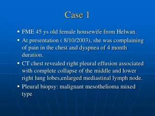
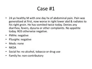
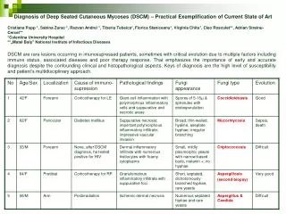
CASE 1. CASE 1. Dystocia. Waiter’s tip sign. http://www.skepticalob.com/2012/10/except-for-the-nerve-damage-the-baby-was-unscathed.html. Upper Lesions of the Brachial Plexus ( Erb-Duchenne Palsy) excessive displacement of the head to the opposite side

E N D
Dystocia Waiter’s tip sign http://www.skepticalob.com/2012/10/except-for-the-nerve-damage-the-baby-was-unscathed.html
Upper Lesions of the Brachial Plexus • (Erb-Duchenne Palsy) • excessive displacement of the head to the opposite side • depression of the shoulder on the same side • in infants during a difficult delivery • in adults after a blow to or fall on the shoulder
Upper Lesions of the Brachial Plexus • (Erb-Duchenne Palsy) • C5 and C6 roots • suprascapular nerve • the nerve to the subclavius • musculocutaneousnerve • axillary nerve • supraspinatus abductor of the shoulder • infraspinatuslateral rotator of the shoulder • subclaviusdepresses the clavicle • biceps brachiisupinator of the forearm, flexor of the elbow, weak flexor of the shoulder • greater part of the brachialis flexor of the elbow • coracobrachialisflexor of the shoulder • deltoid abductor of the shoulder • teresminor lateral rotator of the shoulder Medialrotation Pronation Sensatıonlossoverthelateralsıde of thearm
Dorsalscapularnerve:Levatorscapulae, rhomboids (Retracts (adducts) and elevates scapula) Lateralpectoralnerve: Pectoralismajor (medialrotation, flexionandadduction of thearm) extension Lattisimusdorsi= thoracodorsalnerve
Lower Lesions of the Brachial Plexus (Klumpke Palsy) ulnar and median nerves all the small muscles of the hand Claw(ed) hand hyperextension of metacarpophalangeal joints flexion of the interphalangeal joints
Lower Lesions of the Brachial Plexus (Klumpke Palsy) C8 and T1 roots loss of sensation along the medial side of the arm 8th cervicalnervedamaged + medial side of the forearm, hand, and medial two fingers Foerster (1933)
Mononeuropathies The pattern of distribution of peripheral nerve involvement is very helpful in reaching a diagnosis. Mononeuropathies, especially if an entrapment site, are often an isolated phenomenon, possibly related to pregnancy, DM,thyroid disease or occupation, but importantly may also occur as features of a more generalised disorder, such as hereditary neuropathy with liability to pressure palsies (HNPP) or amyloidosis.
Mononeuropathies occurring outside entrapment sites are more important to investigate fully, especially if vasculitis is suspected as this need careful evaluation for treatment. If the pattern suggests a single nerve or plexus lesion at an unusual site of compression or invasion, such as a radial nerve lesion compressed on a chair in a patient following an overnight binge, or invasion of the brachial plexus with breast malignancy, this is clearly important to detect. PALSY
Focal and multifocal neuropathies Entrapment neuropathy—for example, carpal tunnel syndrome (CTS), ulnar nerve at elbow Myxoedema, acromegaly Amyloidosis Diabetes Hereditary neuropathy with liability to pressure palsies (HNPP A) Vasculitis Multifocal motor neuropathy
Entrapment neuropathies occur when nerves chronically compressed or mechanically injured at specific locations. isolated peripheral nerve injuries occurring at specific locations where a nerve is mechanically constricted in a fibrous or fibro-osseous tunnel or deformed by a fibrous band. In some instances the nerve is injured by chronic direct compression, and in other instances angulation or stretching forces cause mechanical damage to the nerve.
Angulation and stretch injury are important mechanisms of nerve injury for ulnar neuropathies associated with gross deformity of the elbow joint (“tardy ulnar palsy”). Recurrent compression of nerves by external forces may also cause focal nerve injuries such as ulnar neuropathy at the elbow and deep branch lesions of the ulnar nerve in the hand. Although these latter neuropathies do not satisfy the strict definition of “entrapment neuropathies”, they are often considered in a discussion of the topic.
Long Thoracic Nerve Injuries serratus anterior C5, C6, C7 blows to or pressure on the posterior triangle of the neck during the surgical procedure of radical mastectomy • Difficulty in raising the arm above the head • Inferiorborder of scapula not closelyappliedtothechestwall • Protrudeposteriorly • Wingedscapula
CASE 2 Here is the shoulder’s x-ray of the patient! SYMPTOMS loss of skin sensation over the lower half of the deltoid muscle. What is your possible diagnosis?
Axillary Nerve Injuries posterior cord of the brachial plexus (C5 and 6) pressure of a badly adjusted crutch pressing upward into the armpit shoulderdislocationsQuadrangularspace fractures of thesurgicalneck of humerus deltoid and teres minor Loss of skin sensation overthelowerhalf of deltoidregion (lateralpart of thearm) Upperlateralcutaneousnerve of thearm Impairedabduction of theshoulder (theotherone: Supraspinatusonly) Shoulderweakness Difficulty lifting thearmabovethehead
Axillary Nerve Injuries posterior cord of the brachial plexus (C5 and 6) I.M. injections Operations around the shoulder runs transversely under cover of the deltoid at the level of the surgical neck of the humerus
CASE 3 A NIGHT AT E.R. You are taking the history from your patient. Here is what he says (his symptoms) He says he was painting the ceiling and fell., suddenly. He has a terrible pain in his arm. Here is what you find (his clinical findings) Localized pain in his right forearm No sensory loss No wristdrop, the wrist can be extended.
Radial Nerve Injuries commonly damaged in the axilla & in the spiral (radial) groove
Radial Nerve Injuries @ Axilla pressure of the upper end of a badly fitting crutch drunk falling asleep with one arm over the back of a fractures and dislocations of the proximal end of the humerus Motor Triceps, anconeus, and long extensors of the wrist No extension of theelbowjoint, wristjoint, andthefingers Wristdrop(flexion of thewrist) Supinationgoodbrachioradialis, supinatordown, but bicepsbrachii
Radial Nerve Injuries @ Axilla Sensory A small loss of skin sensation down the posterior surface of the lower part of the arm down a narrow strip on the back of the forearm A variable area of sensory loss on the lateral part of the dorsum of the hand on the dorsal surface of the roots of the lateral 3 ½ fingers Trophic Changes Slight
Radial Nerve Injuries @ Spiral Groove of Humerus At the time of fracture of the shaft of the humerus Followingtheformation of the callus Pressure of the back of the arm on the edge of the operating table Prolonged application of a tourniquet to the arm in a person with a slender triceps temporary radial palsy
Radial Nerve Injuries @ Spiral Groove of Humerus mostcommonly @ distalpart of thegroove Motor Inabilityto extend the wrist &fingers Wristdrop Sensory A variable small area of anesthesia dorsal surface of the hand dorsal surface of roots of lateral 3 ½ fingers Trophicchanges Very slight or absent
RadialTunnel potential space located anterior to the proximal radius starting from the level of the humeroradial joint extending past the proximal edge of the supinator posterior interosseus nerve The radial nerve bifurcates into deep and superficial branches anterior to the lateral epicondyle of the humerus, between the brachialis and the brachioradialis, in the lateral border of the cubital fossa. After passing through the two heads of the supinator muscle, the deep branch becomes the posterior interosseous nerve.
Radial tunnel syndrome DIAGNOSIS Radialnerve Lateralpart of theelbow, radialtunnelbelowthesupinator Tenderness and pain @ lateral side of the elbow ANATOMY
TennisElbow(Lateralepicondylitis) sudden and often repeated use of the forearm extensor muscles previously been much used extensor carpi radialisbrevis Tenderness and pain @ lateral side of the elbow Pain on wrist extension, pain when shaking hands, and frequently a weakened grip. In tennis elbow, the tenderness ismostly right where the tendon attaches to the lateral epicondyle of the elbow. Inradial tunnel syndrome, the place that ismost tender is about two inches furtherdown the arm, right over where theradial nerve goes into the supinatormuscle.
Deepbranch of theRadialNerveInjuries Motor nerve Extensormuscles @ posteriorcompartment of theforearm fractures of the proximal end of the radius dislocation of the radial head SupinatorIntact Extensor carpi radialislongus No wristdrop No sensoryloss
SuperficialRadialNerveInjuries a variable small area of anesthesia over the dorsum of the hand dorsal surface of the roots of the lateral 3 ½ fingers
Musculocutaneous Nerve Injuries Rarelyinjured (protectedposition) • Weakflexion @ shoulderjoint • Flexion of theforearm @ elbowbyremainder of brachialis + flexors of forearm • Weaksupinationsupinatorradialnerve • Sensory loss along the lateral side of the forearm • lateral cutaneous nerve of the forearm
Median Nerve Injuries occasionally in the elbow region supracondylar fractures of the humerus most commonly by stab wounds or broken glass just proximal to the flexor retinaculum
Injuries to the Median Nerve @ the Elbow Motor pronator & flexormuscles of forearm (EXCEPT?) thenarmuscles Forearm in supineposition- Weakwristflexion-accompaniedbyadduction No flexion @ interphalangealjoints of index & middlefingers Weakflexion @ metacarpophalangealjoints –interossei- Middle & index fingers remain straight (extended) FIST POPE’S BLESSING
Injuries to the Median Nerve @ the Elbow Motor No Flexion of the terminal phalanx of the thumb Thenareminenceflattened Thumblaterallyrotated & adducted APE HAND DEFORMITY
Injuries to the Median Nerve @ the Elbow Sensory Lost skin sensation @lateralhalforless of thepalm of thehand palmaraspect of lateral 3 ½ fingers distal part of dorsal surfaces of lateral 3 ½ fingers
Injuries to the Median Nerve @ the Elbow Vasomotorchanges Skin areaaffectedwarmer & drier Arteriolar dilatation and absence of sweating / loss of sympathetic control Trophicchanges Chroniccases dry and scaly skin nails crack easily atrophy of the pulp of the fingers
Injuries to the Median Nerve @ the Wrist Motor Thenarmuscles & firsttwolumbricals Thenareminenceflattened Thumblaterallyrotatedandadducted Ape-likehand No opposition of thethumb MAKE A FIST, SLOWLY Index & middlefingerslagbehindthe ring & littlefingers
CASE 4 Dr. Eda, a gynecologist sends her patient , 25 yearsoldpregnantfemale , a cashier at a mall in downtown @ theend of her firsttrimesterto Dr. Süleyman who is an internist An expertfrom Dr. Eda’srequest of consultationletterfor Dr. Süleyman FreeT4 0.4 ng/dl 0.8 – 2.4 ng/dl Hypothyroidism?
CASE 4 Dr. Süleyman examinesthepatient as a consultinginternist, Findsthefollowingduring his examination: Symptoms of depression Increasedsensitivity to cold Poormuscletone Burningsensations in the thumb & indexfinger Theanxiouspatientasks: What is wrongwith me, doctor? Dr. Süleyman replieswith a tone of affection in his voice: «Youarepregnantandyourthyroidglanddoes not workefficientlyandplusyouhave …………………..X………………» What is X?
Carpal tunnel syndrome (CTS) is a frequent diagnosis considered in such patients and is the most frequent entrapment neuropathy. Approximately 1.6% of adults describe symptoms consistent with CTS. Clinically, thesyndromeconsists of a burningpainor “pinsandneedles” alongthedistribution of themediannervetothelateralthreeand a halffingersandweakness of thethenarmuscles. It is producedbycompression of themediannervewithinthetunnel.The syndrome can often be treated effectively with splinting and avoidance of repetitive motions and awkward wrist positions; however, carpal tunnel release is ultimately performed in 25% to 50% of these patients. Solomon DH, Katz JN, Bohn R, Mogun H, Avorn J. Nonoccupational risk factorsforcarpaltunnelsyndrome. J Gen InternMed. 1999 May;14(5):310-4. Pregnancyexperience CTS duetohormonalchanges (highprogesteronelevels) andwaterretention, which is commonduringpregnancy Duringhypothyroidismandpregnancyfluid is retained in tissues, whichswellsthetenosynovium. Obesity also increases the risk of CTS: individuals classified as obese (BMI > 29) are 2.5 times more likely than slender individuals (BMI < 20) to be diagnosed with CTS. CTS is alsowork-related, occupationaldiseaseandassociatedwithoveruse. Bonfiglioli R, Venturi S, Graziosi F, Fiorentini C, Mattioli S. [Carpaltunnelsyndromeamongsupermarketcashiers]. G ItalMed Lav Ergon. 2005 Jan-Mar;27(1):106-11. [Article in Italian]
Carpal Tunnel Syndrome MOST COMMON PERIPHERAL NERVE INJURY IN THE UPPER LIMB
Carpal Tunnel Syndrome Burning pain or “pins and needles” along the distribution of the median nerve tothelateral 3 ½ fingers Weakness of thenar muscles No paresthesiaoverthethenareminence palmar cutaneous branch of the median nerve
Ulnar Nerve Injuries most commonly injured @ @ elbow where it lies behind the medial epicondyleusuallyassociatedwithfracture @ wrist where it lies with the ulnar artery in front of the flexor retinaculum.
Injuries to the Ulnar Nerve @ theElbow CUBITAL TUNNEL SYNDROME (2ND mostcommon) Motor Flexor carpi ulnaris& medial half of flexor digitorumprofundus ring & littlefingers No flexion of the terminal phalanges of the ring & littlefingers Flexion of wrist = abductionparalysis of flexorcarpiulnaris medial border of the front of the forearmflattned/wasted Allthesmallmuscles of thehandparalyzedEXCEPT ? fibro-osseus tunnel between the medial epicondyle and flexor carpi ulnaris
Injuries to the Ulnar Nerve @ theElbow Motor Extensordigitorum can abduct the fingers to a small extent when metacarpophalangeal joints are hyperextended Impossible toadductthethumbadductorpollicisparalyzed Froment’ssign Grip a piece of paperbetweenthethumbandindexfingers Froment sign: The patient is asked to hold the paper between the thumb and index finger. (A) With the intact ulnar nerve, the patient is able to make use of the adductor pollicis. ( B) When the ulnar nerve is deficient, the patient compensates for the denervated adductor by using the flexor pollicis longus (median nerve innervated).
Injuries to the Ulnar Nerve @the Elbow Motor 2 mediallumbricals & interosseiHyperextendedmetacarpophalangeal joints Flexedinterphalangeal joints fourth &fifth fingers “claw” deformity main en griffe
Injuries to the Ulnar Nerve @ theElbow Motor Flattening of hypothenareminence Loss of theconvexcurvetothemedialborder of thehand Hollowingbetweenmetacarpalbones @ dorsum of thehand wasting of dorsalinterossei
Injuries to the Ulnar Nerve @ theElbow Sensory Loss of skin sensation anterior & posterior surfaces of medial 1/3 of the hand medial 1 ½ fingers Vasomotor Changes warmer and drier skin area arteriolar dilatation and absence of sweating /loss of sympathetic control
Injuries to the Ulnar Nerve @ theWrist Motor Small handmusclesparalyzed, wasted – EXCEPT 3 thenar @ first 2 lumbricals Clawhand Moreobvious Flexordigitorumprofundusintact Markedflexion of the terminal phalanges Ulnarparadox Higherlesion Lessobviousclawdeformity Moreproximalinjury Lessclaw