Download
1 / 37
380 likes | 662 Views
Female pelvis . Fetus as the object of labor . Obstetric terminology METHODS OF EXAMINATION doc. Stelmakh O.Y. Female pelvis.

E N D
Female pelvis. Fetus as the object of labor. Obstetric terminology METHODS OF EXAMINATION doc. Stelmakh O.Y.
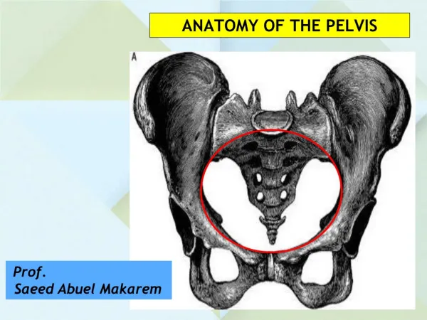
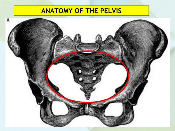
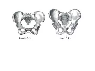
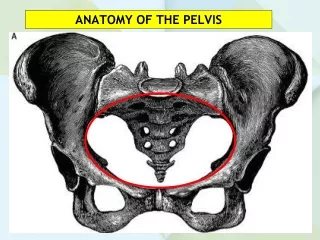
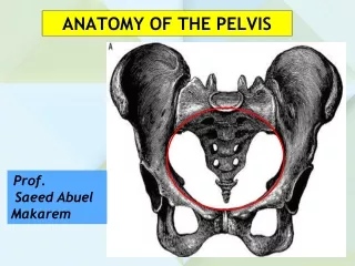
Female pelvis • Birth canal is divided into bone and soft parts tobone belongs small pelvis and to soft - cervix, vagina,muscle-fascial system of the pelvic floor.Bone part of the pelvisFemale pelvis with obstetric considerations are divided into two sections: the large andsmall pelvis. The boundary between them passes through an unmarked line (linea innominata). Large pelvis bounded on the sides of the iliac wings,back - the spine. Small pelvis formed in frontbranches of the pubic bones and symphysis on each side - Part bonesconstitute acetabular and ischial bones, behind -the sacrum and coccyx.During childbirth small pelvis as dense bone tunnel limits anddetermines the size, shape and direction of the birth canal, which fetuspasses, and has to adapt by changing their own configuration.
The main dimensions of the pelvis • Of all the methods of the pelvis examination is essential it measurement.Most internal pelvic sizes available for measurement, sousually measured by its external dimensions and evaluate them aboutinternal.Measurement of pelvic spend by pelviometr.Usually measured four basic dimensions of the pelvis: three transverse and one direct.
The main dimensions of the pelvis • Distantia spinarum - the distance between the upper anterior iliac spine bones. This size is 25-26 cm;Distantia cristarum - the distance between the most distant points of wings iliac bone. On average it is 28 cmDistantia trochanterica - the distance between the trochanter major of hipbones. This size is 31 cm
The main dimensions of the pelvis • external conjugate – external size of pelvis. End of pelviometr set onmiddle of the upper margin of symphysis, the other end is over the sacral fossa contained between fifth lumbar vertebra and the beginning of the first sacral vertebra. External conjugate is20 cm
The planes of the pelvis and their dimensions • In obstetric practice are important dimensions of the pelvis, fromwhich depends on the course and outcomes for both mother and fetus. But most sizes pelvis can not be measured directly.Great pelvis for childbirth substantial does not matter, but in itssize may indirectly informates about the form and size of the pelvis.Pelvic cavity is the space between the walls, which the top and bottom limited inlet and outlet planes of the pelvis. It looks like a cylinder. In pelvic cavity are four planes:inlet, greatest dimention,narrow part (midpelvis) and outlet.
The planes of the pelvis and their dimensions • The plane of the inlet of a small pelvis bounded behind promontory of sacral bone, laterally- arcuate lines iliac bone, antioriorly- uppermargin of the pubic bone and symphysis.
Small pelvis. The planes and the size of the pelvis • At the pelvic inlet there are4 sizes: antirior-postirior, transvers and two oblique. Direct size - distance from sacral promontory to the point that most appear on the inner surface of the upper margin of the pubic symphysis - is true or obstetric conjugate (conjugata vera), which is 11 cm away from promontory the to the middle of the upper part of symphysis anatomical conjugate to 0.3-0.5 cm greater than the obstetric .
Small pelvis. The planes and the size of the pelvis • Dimensions plane of the pelvic inlet 1 – antirior-posterior size, 2 - transverse size, 3 - left oblique size, 4 - right oblique sizeThe transverse size of the distance between the inniminata lines is 13 cm • Two oblique, right and left, which have 12 cm Right oblique size goes from the right iliac-sacral junction to the left eminentia iliopubis. Left oblique size- from the left iliacsacrum junction to the right eminentia iliopubis.
Small pelvis. The planes and the size of the pelvis • Plane of widest part of the pelvic cavity has the following limits:front - a middle inner surface of the pubic symphysis, laterally - mid acetabulum hollow, back - the place II and III sacral vertebrae. Direct and transverse dimensions of the plane are equal to 12,5 cm
Small pelvis. The planes and the size of the pelvis • The plane of the narrow part of the pelvic cavity is limited to the lower edge of the front pubic symphysis, laterally – ishial spine behind - sacro-coccygeal joints. Direct size is 11-11,5 cm transvers - 10.5 cm
Small pelvis. The planes and the size of the pelvis • Plane of pelvic outlet has the following borders: anteriorly- lower margin of the pubic symphysis, laterally –tuber ischii, posteriorly- the tip of the coccyx. Direct size - 9.5 cm, and the transvers - 11.5 cmThe line connecting the centers of all direct sizes of pelvis, called the main axis of the pelvis
Measuring the size of the pelvis • Diagonal conjugate measured during vaginal examination from the lower margin of pubis to promontory. Normally, this distance is 12,5-13 cm for the determination of the real conjugates of bias should be deducted 1.5-2 cmIndex Soloviev - average circumference of wrist is 14 cmRhombus of Michaelis - upper angle contained hollow under the spinous processes of the 5 lumbar vertebrae Lateral angles correspond posterior-superior iliac spine, lower- top sacrum. In women with a normal pelvis it has the correct form, approaching the square, its dimensions are 10.11 cm, height of the upper triangle 3-3.5 cm
Fetus as the object of labor • From all parts of the mature fetus most interesting head, because of the following reasons: 1) head has the big circumference and dense part of thefetus, which can withstand the greatest resistance from the birth canal and puts most pressure on them that determines the outcome of labor, 2) depending on the density and mobility of the cranial bones is greatly damage the birth canal of the mother and the fetus, and 3) the head of the fetus has a large number of cognitive items, which helps in diagnosing insertion and promotion in the bones of the pelvis.At the head of the fetus can distinguish two parts (Fig. 1): a relatively small front: lower jaw (1), maxilla (2) and very voluminous - brain. The latter consists of seven bones: two frontal (3), two parietal(4), one occipital (5), two temporal (6).
Fetus as the object of labor • Sutures and fontanelles skull newborn (seen from above):1 - frontal suture, 2 - coronal suture 3 - sagittal (sagittal) suture 4 - occipital suture, 5 - Small fontanel 6 - large fontanelAll bony parts are interconnected fibrous membranes, allowing the process of childbirth crossbones go one after another, thus reducing the size of the head
Fetus as the object of labor • These fibrous membrane called sutures There are the following joints: 1) frontal sutura (sutura frontalis); 2) coronal suture (sutura coronaria), connecting on each side of the frontal and parietal bones, and 3) sagittal (sagittal) suture (sutura sagittalis), combining two parietal bone, 4) or lyambdoid occipital suture (sutura lambdoidea), connecting occipital bone with the parietal, 5) temporal suture (sutura temporalis), connecting on each side of the parietal temporal bone (mostly).
Fetus as the object of labor • Fibrous membrane at the intersections of joints called fontanelles. There are two main fontanel and two pairs of secondary. The main fontanelles include large and small fontanel Large fontanel (fonticulus magnus s. Bregmaticus) located at the intersection of coronary, windshield and sagital suture and a diamond shape. Acute angle of the diamond sent to the forehead, and - back. It is easily determined by finger. Small (5) fontanel is located at the intersection sagital and occipital sutures. Unlike large, small fontanel poorly defined because it is already ripe fetus filled bone.
Fetus as the object of labor • At the head term fetus can distinguish the following dimensions and perimeter :1. vertical size (diameter sublinguo-bregmatica) distance from the hyoid bone to the middle of the large fontanel, is equal to 9.5 cm on the contours of the head, measured through these points vertical size, (circumferentia sublinguo-bregmatica) - 32 cm2. Large oblique size (diameter mento-occipitalis) - from the chin to the farthest point of the neck, length - 13 cm from the perimeter (circumferentia mento-occipitalis) - 41 cm
Fetus as the object of labor • 8. Direct size (diameter fronto-occipitalis) - from the nose to the occipital hill, length - 12 cm by 34 cm contours of equal • 7. Average oblique size (diameter suboccipito-frontalis) - from suboccipital fossa to the anterior border of the scalp, length of 10 cm, and contours (circumferentia suboccipito-frontalis) - 33 cm • 6. Small oblique size (diameter suboccipito-bregmatica) - from the middle suboccipital fossa large fontanel, length - 9.5 cm, and circumference suboccipito-bregmatica - 32 cm
Fetus as the object of labor • 7. Small transverse size (diameter bitemporalis) - the distance between the most distant points of the coronal suture, - 8 cm8. Large transverse size (diameter biparietalis) - the distance between the parietal bones, length - 9.5 cm Circumferences of shoulders is 34cm (12cm), circumferences of pelvic part is 28cm (9,5cm).
Fetus as the object of labor • The transverse size of the buttocks (distantia bisiliacalis) length and 9.5 cm in perimeter: an incomplete presentation buttocks - 32 cm , with full foot previa - 28 cm , with full presentation buttocks - 34 cm . 11. Contours (in cm) newborn body in full foot presentation: shoulder with handles (34) buttocks (28). 12. Contours (in cm) newborn body in complete breech presentation: shoulder (34); buttocks with legs (34).
Obstetric terminology • definitions of "fetal lie, "position", "view", "presentation"Accurate knowledge of the position of the fetus in the uterus, is of great importance for practical obstetrics. It is achieved by the examination of women in late pregnancy, when you can set fetus habitus, itslie, presentation, position and variety.
Obstetric terminology • Attitude of fetus (habitus) - is the ratio of the limbs of the fetus and the head to his body. In the most favorable habitus - curved spine, resulting in back arched outwards, head bent, chin close to the chest. The legs are flexed at the hip and knee joints, intersect and pinned to the lower abdomen. Handles are flexed at the elbows and intersect on his chest.
Obstetric terminology • Fetal lie (situs) - is the ratio of the axis of the fetus to the axis of the uterus. The axis of the fetus - a line that passes through the neck and buttocks. Can meet the following options for the of the fetus lie : fetal axis coincides with the uterus - longitudinal lie (situs longitudinalis) - occurs in 99% of cases; fetal axis intersects the uterus - transverse (situs transversus) or oblique fetal lie (situs obliguus).
Obstetric terminology • Position of the fetus (positio) - is the ratio of the fetus back to the left (I position 2/3 cases) or right (second position, 1/3 of cases) the uterine wall. In transverse position of the fetus position is determined by the placement head.Type of position (visus) - the ratio of the fetus back to the front or back wall of the uterus. In the front form the back of the fetus is facing the front wall of the uterus, at the back of the form - to the back wall of the uterus.Fetal presentation (presentatio) - is the ratio of the lowest placed a large part of the fetus to enter the pelvis (main or breech).
Fetal head station • -2 (fixed to pelvic inlet) • -1(small segment of fetal head in pelvic inlet) • 0 (large segment of fetal head in pelvic inlet) • +1 (fetal head in plane of greatest dimension) • +2 (fetal head in plane of least dimension) • +3 (fetal head in the pelvic outlet
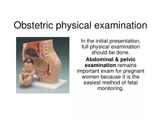
METHODS OF EXAMINATION • Anamnesis womenWhen taking history clarifies the following issues: 1) Passport part: last name, first name, age, pregnancy, occupation, address, phone, and 2) carried diseases: childhood, adulthood, pregnancy, and 3) family history, and 4) working and living conditions, and 5) special history: menstrual, sexual, reproductive and secretory functions, 6) the occurrence of pregnancy.
METHODS OF EXAMINATION • Overview pregnantDuring the general examination pay attention to height, body structure, completeness, condition of skin, shape of the abdomen, development of breasts and nipples, pubic hair growth
METHODS OF EXAMINATION • External measurementIn order to have an idea about the size of the fetus, measure the size of the abdomen measuring tape: sight abdoman. In the supine position at the navel at the end of pregnancy, this woman is 110 cm and the height stoyanyya uterus above the pubis - 37 cm to determine the mass of fetal abdominal perimeter must be multiplied by the height of standing uterus (110 cm x 37 cm = 4070 g).
METHODS OF EXAMINATION • External examinationExternal examination -Leopold-Levitsky manuvers:The first external method • Obstetrician sits to the right of the pregnant woman, facing her. Palmar surface of the hand puts the fundus of the uterus, is trying to bring together the nail phalanges. This reception determine the height of standing and form the uterus (normal, saddle, two-horned), the part of fetus that is at its fundus .
METHODS OF EXAMINATION • The second external method.. The doctor pulls the two arms of the uterus on the side wall of the abdomen and one by one, then another, then another hand, carries palpation. If it finds one side smooth, wide, curved surface - back (left - the first position to the right - the second position), which is facing forward (front view) or back (posterior view.
METHODS OF EXAMINATION • The third external method . Obstetrician palm and thumb and other four fingers of his right hand covers and squeezes the fetus placed above the entrance to a smallpelvis, attempts to displace it to the right or left side, verifying the presence or absence of symptoms ballotment. If above the pubic joints palpated soft and broad, it can be suspected breech presentation, and if solid and rounded -cefalic .
METHODS OF EXAMINATION • Fourth external method is performed as follows: Obstetrician stands face to legs bent at the knees pregnant tips and palmar surfaces of the fingers of both hands gently and gradually slides along the side of the head down, gets between her and the plane door in a small bowl and returns back up, checking the results. In transverse position of the fetus peredlezhascha part on the pubic joints not palpable and fingers freely agree among themselves high above the vagina. When cephalic this method allows to determine the place of accommodation. If peredlezhascha part above the entrance to the small pelvis, the fingers of both hands freely converge under it, and when returning back - apart
METHODS OF EXAMINATION • Vaginal examinationVaginal examination is required in the following cases: the first - at the time of admission to the hospital pregnant, the second - after the discharge of amniotic fluid or early labor activity and the third - when changing obstetric situation; fourth - early in the second stage of labor Vaginal examination provides information on the status of the genital tract before birth, there exostosis, bone tumors, deformities of the pelvis, dynamic opening of the cervix, the presence of amniotic mamrane, the mechanism of insertion and passage of the birth canal presenting part.
METHODS OF EXAMINATION • Ultrasonic dating of the pregnancy and an ultrasonic fetal survey to detect gross abnormalities have been recommended in some clinics as a routine part of early prenatal care. Routine ultrasonography is most cost – effective in patients in whom the date of the last menstrual period is uncertain and in patients with a family history of congenital anomalies. Considerable individualization should be exercised in making the decision to order this evaluation. If ultrasonography is performed, it is most informative between 11-13 and18-20 weeks.
METHODS OF EXAMINATION • Auscultation. In cephalic presentation, the point of maximal intensity of fetal heart sounds is usually midway between the maternal umbilicus and the anterior-superior spine of her ilium.