Download
1 / 67
670 likes | 688 Views
Explore the composition of blood, its physical characteristics, functions, and the role of plasma and formed elements. Learn about erythrocytes, leukocytes, platelets, and the process of hematopoiesis.

E N D
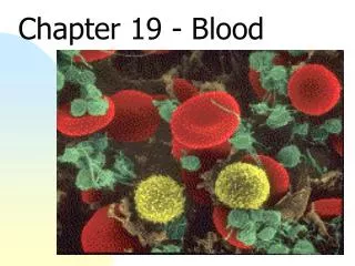
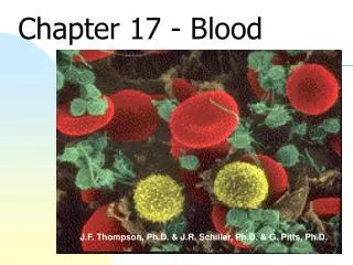
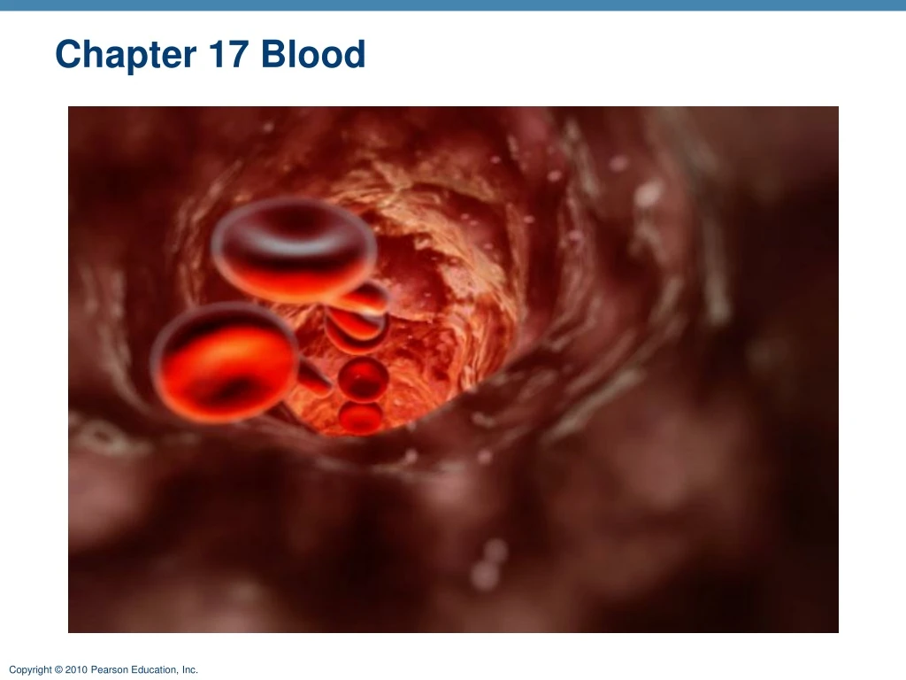
Blood Composition • Blood: a fluid connective tissue composed of • Plasma • Formed elements • Erythrocytes (red blood cells, or RBCs) • Leukocytes (white blood cells, or WBCs) • Platelets
Blood Composition • Hematocrit • Percent of blood volume that is RBCs • 47% ± 5% for males • 42% ± 5% for females
Physical Characteristics and Volume • Sticky, opaque fluid • Color scarlet to dark red • pH 7.35–7.45 • 38C • ~8% of body weight • Average volume: 5–6 L for males, and 4–5 L for females
Functions of Blood • Distribution of • O2 and nutrients to body cells • Metabolic wastes to the lungs and kidneys for elimination • Hormones from endocrine organs to target organs
Functions of Blood • Regulation of • Body temperature by absorbing and distributing heat • Normal pH using buffers • Adequate fluid volume in the circulatory system
Functions of Blood • Protection against • Blood loss • Plasma proteins and platelets initiate clot formation • Infection • Antibodies • Complement proteins • WBCs defend against foreign invaders
Blood Plasma • 90% water • Proteins are mostly produced by the liver • 60% albumin • 36% globulins • 4% fibrinogen
Blood Plasma • Nitrogenous by-products of metabolism—lactic acid, urea, creatinine • Nutrients—glucose, carbohydrates, amino acids • Electrolytes—Na+, K+, Ca2+, Cl–, HCO3– • Respiratory gases—O2 and CO2 • Hormones
Formed Elements • Only WBCs are complete cells • RBCs have no nuclei or organelles • Platelets are cell fragments • Most formed elements survive in the bloodstream for only a few days • Most blood cells originate in bone marrow and do not divide
Platelets Erythrocytes Monocyte Neutrophils Lymphocyte Figure 17.2
Erythrocytes • Biconcave discs, anucleate, essentially no organelles • Filled with hemoglobin (Hb) • Major factor contributing to blood viscosity
Erythrocytes • Structural characteristics contribute to gas transport • Biconcave shape—huge surface area relative to volume • >97% hemoglobin (not counting water) • No mitochondria; ATP production is anaerobic; no O2 is used in generation of ATP
Hemoglobin (Hb) • O2 loading in the lungs • Produces oxyhemoglobin (ruby red) • O2 unloading in the tissues • Produces deoxyhemoglobin or reduced hemoglobin (dark red) • CO2 loading in the tissues • Produces carbaminohemoglobin (carries 20% of CO2 in the blood)
Hematopoiesis • Hematopoiesis (hemopoiesis): blood cell formation • Occurs in red bone marrow of axial skeleton, girdles and proximal epiphyses of humerus and femur • Hemocytoblasts (hematopoietic stem cells) • Give rise to all formed elements • Hormones and growth factors push the cell toward a specific pathway of blood cell development
Erythropoiesis • Erythropoiesis: red blood cell production • A hemocytoblast is transformed into a proerythroblast • Proerythroblasts develop into early erythroblasts
Regulation of Erythropoiesis • Too few RBCs leads to tissue hypoxia • Too many RBCs increases blood viscosity • Balance between RBC production and destruction depends on • Hormonal controls (EPO) • Adequate supplies of iron, amino acids, and B vitamins
Fate and Destruction of Erythrocytes • Life span: 100–120 days • Old RBCs become fragile, and Hb begins to degenerate • Macrophages engulf dying RBCs in the spleen
Fate and Destruction of Erythrocytes • Heme and globin are separated • Iron is salvaged for reuse • Heme is degraded to yellow the pigment bilirubin • Liver secretes bilirubin (in bile) into the intestines • Degraded pigment leaves the body in feces as stercobilin • Globin is metabolized into amino acids
Erythrocyte Disorders • Anemia: blood has abnormally low O2-carrying capacity • A sign rather than a disease itself • Blood O2 levels cannot support normal metabolism • Accompanied by fatigue, paleness, shortness of breath, and chills • Three causes • Low RBC count • Low Hg • Abnormal Hg
Causes of Anemia • Sickle-cell anemia • Defective gene codes for abnormal hemoglobin (HbS) • Causes RBCs to become sickle shaped in low-oxygen situations
Erythrocyte Disorders • Polycythemia: excess of RBCs that increase blood viscosity • Results from: • Polycythemia vera—bone marrow cancer • Secondary polycythemia—when less O2 is available (high altitude) or when EPO production increases • Blood doping-artificially induced polycythemia
Leukocytes • White blood cells (WBC’s) • <1% of total blood volume • Can leave capillaries via diapedesis • Move through tissue spaces by ameboid motion and positive chemotaxis • Leukocytosis: WBC count over 11,000/mm3 • Normal response to bacterial or viral invasion
Granulocytes • Granulocytes: neutrophils, eosinophils, and basophils • Cytoplasmic granules stain specifically with Wright’s stain • Larger and shorter-lived than RBCs • Lobed nuclei • Phagocytic
Neutrophils • Most numerous WBCs • Polymorphonuclear leukocytes (PMNs) • Give the cytoplasm a lilac color • Granules contain hydrolytic enzymes or defensins • Very phagocytic—“bacteria slayers”
Eosinophils • Red-staining, bilobed nuclei • Digest parasitic worms that are too large to be phagocytized • Modulators of the immune response
Basophils • Rarest WBCs • Large, purplish-black (basophilic) granules contain histamine • Are functionally similar to mast cells
Agranulocytes • Agranulocytes: lymphocytes and monocytes • Lack visible cytoplasmic granules • Have spherical or kidney-shaped nuclei
Lymphocytes • Large, dark-purple, circular nuclei with a thin rim of blue cytoplasm • Mostly in lymphoid tissue; few circulate in the blood • Crucial to immunity • Two types • T cells - virus-infected cells and tumor cells • B cells - produce plasma cells, which produce antibodies
Monocytes • The largest leukocytes • Abundant pale-blue cytoplasm • Dark purple-staining, U- or kidney-shaped nuclei • Become macrophages
Leukopoiesis • Production of WBCs • Stimulated by chemical messengers from bone marrow and mature WBCs • Interleukins (e.g., IL-1, IL-2) • Colony-stimulating factors (CSFs) named for the WBC type they stimulate (e.g., granulocyte-CSF = granulocytes) • All leukocytes originate from hemocytoblasts
Leukocyte Disorders • Leukopenia • Abnormally low WBC count—drug induced • Leukemias • Cancerous conditions involving WBCs • Named according to the abnormal WBC clone involved • Myelocytic leukemia involves myeloblasts • Lymphocytic leukemia involves lymphocytes • Acute leukemia involves blast-type cells and primarily affects children • Chronic leukemia is more prevalent in older people
Leukemia • Bone marrow totally occupied with cancerous leukocytes • Immature nonfunctional WBCs in the bloodstream • Death caused by internal hemorrhage, anemia, and infections • Treatments include irradiation, antileukemic drugs, and stem cell transplants
Platelets • Not cells, but rather cell fragments of megakaryocytes • Formation is regulated by thrombopoietin • Blue-staining outer region, purple granules • Granules contain serotonin, Ca2+, enzymes, ADP, and platelet-derived growth factor (PDGF)
Hemostasis • Fast series of reactions for stoppage of bleeding • Vascular spasm • Platelet plug formation • Coagulation (blood clotting)
Vascular Spasm • Vasoconstriction of damaged blood vessel • Triggers • Direct injury • Chemicals released by endothelial cells and platelets • Pain reflexes
Platelet Plug Formation • Positive feedback cycle • At site of blood vessel injury, platelets • Stick to exposed collagen fibers not to each other • Swell, become spiked and sticky, and release chemical messengers • ADP - causes more platelets to stick and release their contents • Serotonin and thromboxane A2 - enhance vascular spasm and more platelet aggregation
Coagulation • A set of reactions in which blood is transformed from a liquid to a gel • Reinforces the platelet plug with fibrin threads • Three phases of coagulation • Prothrombin activator is formed • Prothrombin is converted into thrombin • Thrombin catalyzes the joining of fibrinogen to form a fibrin mesh
Phase 1 Intrinsic pathway Extrinsic pathway Tissue cell trauma exposes blood to Vessel endothelium ruptures, exposing underlying tissues (e.g., collagen) Platelets cling and their surfaces provide sites for mobilization of factors Tissue factor (TF) XII Ca2+ XIIa VII XI XIa VIIa Ca2+ IX IXa PF3 released by aggregated platelets VIII VIIIa TF/VIIacomplex IXa/VIIIacomplex X Xa Ca2+ V PF3 Va Prothrombin activator Figure 17.14 (1 of 2)
Coagulation Phase 1: Two Pathways to Prothrombin Activator • Initiated by either the intrinsic or extrinsic pathway (usually both) • Triggered by tissue-damaging events • Involves a series of procoagulants • Each pathway cascades toward factor X • Factor X complexes with Ca2+, PF3, and factor V to form prothrombin activator
Coagulation Phase 2: Pathway to Thrombin • Prothrombin activator catalyzes the transformation of prothrombin to the active enzyme thrombin
Coagulation Phase 3: Common Pathway to the Fibrin Mesh • Thrombin converts soluble fibrinogen into fibrin • Fibrin strands form the structural basis of a clot • Fibrin causes plasma to become a gel-like trap for formed elements • Thrombin (with Ca2+) activates factor XIII which: • Cross-links fibrin • Strengthens and stabilizes the clot
Clot Retraction • Actin and myosin in platelets contract within 30–60 minutes • Platelets pull on the fibrin strands, squeezing serum from the clot
Fibrinolysis • Begins within two days • Plasminogen in clot is converted to plasmin by tissue plasminogen activator (tPA), factor XII and thrombin • Plasmin is a fibrin-digesting enzyme
Factors Limiting Clot Growth or Formation • Two homeostatic mechanisms prevent clots from becoming large • Swift removal and dilution of clotting factors • Inhibition of activated clotting factors • Heparin – blocks the intrinsic pathway • Warfarin (Coumadin) – inhibits Vitamin K binding to clotting factors
Factors Preventing Undesirable Clotting • Platelet adhesion is prevented by • Smooth endothelial lining of blood vessels • Antithrombic substances nitric oxide and prostacyclin secreted by endothelial cells • Vitamin E quinine, which acts as a potent anticoagulant
Disorders of Hemostasis • Thromboembolytic disorders: undesirable clot formation • Bleeding disorders: abnormalities that prevent normal clot formation
Thromboembolytic Conditions • Thrombus: clot that develops and persists in an unbroken blood vessel • May block circulation, leading to tissue death • Embolus: a thrombus freely floating in the blood stream • Pulmonary emboli impair the ability of the body to obtain oxygen • Cerebral emboli can cause strokes
Disseminated Intravascular Coagulation (DIC) • Widespread clotting blocks intact blood vessels • Severe bleeding occurs because residual blood unable to clot • Most common in pregnancy, septicemia, or incompatible blood transfusions