Chapter 10 Blood
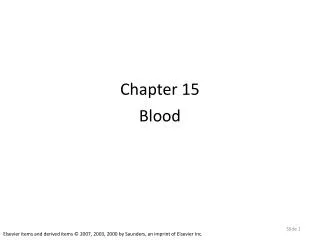
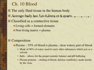
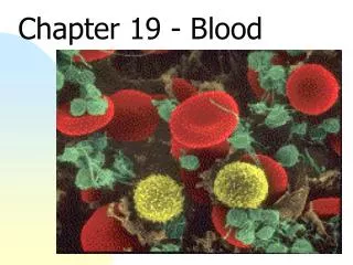
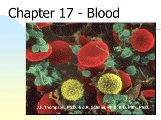
Chapter 10 Blood. Blood. The only fluid tissue in the human body Classified as a connective tissue Living cells = formed elements Non-living matrix = plasma Hematocrit (HCT) Blood spun in centrifuge Rbc- 45% Buffy coat – 1% Plasma – 55%. Blood. Figure 10.1.

Chapter 10 Blood
E N D
Presentation Transcript
Blood • The only fluid tissue in the human body • Classified as a connective tissue • Living cells = formed elements • Non-living matrix = plasma • Hematocrit (HCT) • Blood spun in centrifuge • Rbc- 45% • Buffy coat – 1% • Plasma – 55%
Blood Figure 10.1
Physical Characteristics of Blood • Color range • Oxygen-rich blood is scarlet red • Oxygen-poor blood is dull red • pH must remain between 7.35–7.45 • Regulated by respiratory & urinary systems • Blood temperature is slightly higher than body temperature - 38°C (100.4°F)
Sticky, opaque, metallic taste • Viscous • = 8% of body weight; 5-6L
Blood Plasma • Composed of approximately 90 percent water • Includes many dissolved substances (>100) • Nutrients • Salts (metal ions) • Respiratory gases • Hormones • Proteins • Waste products • Straw colored
Plasma Proteins • Albumin – regulates osmotic pressure • Keeps water in blood • Made in liver • Clotting proteins – help to stem blood loss when a blood vessel is injured • Antibodies – help protect the body from antigens & pathogens
Formed Elements • Erythrocytes = red blood cells • Leukocytes = white blood cells • Platelets = cell fragments
Photomicrograph of a Blood Smear Figure 10.2
Erythrocytes (Red Blood Cells) • The main function is to carry oxygen • Anatomy of circulating erythrocytes • Biconcave disks – large surface area relative to volume • Essentially bags of hemoglobin • Anucleate (no nucleus) • Contain very few organelles • No mitochondria • Anaerobic respiration so don’t use up oxygen • Outnumber white blood cells 1000:1 • 4-6 million/mm3
Hemoglobin • Iron-containing protein • Binds strongly, but reversibly, to oxygen • Each hemoglobin molecule has four oxygen binding sites • Each erythrocyte has 250 million hemoglobin molecules • Can carry 1 billion molecules of oxygen • Normal – 12-18g/dl
Anemia - ↓ # of rbcs or ↓ hgb or abnormal hgb • Sickle cell anemia: • Hgb S due to abnormal amino acid • Rbcs sickle & rupture & clog vessels • Don’t carry enough oxygen • Makes immune from malaria • Sickle cell trait: no symptoms
Polycythemia: • ↑ rbcs • Polycythemia vera – bone marrow cancer or living at high altitudes • ↑ blood viscosity • Iron deficiency anemia: • Common in females
Leukocytes (White Blood Cells) • Crucial in the body’s defense against disease • These are complete cells, with a nucleus and organelles • Able to move into and out of blood vessels (diapedesis) • Can move by ameboid motion • Can respond to chemicals released by damaged tissues – positive chemotaxis
Leukocyte Levels in the Blood • Normal levels are between 4,000 and 11,000 cells per cubic millimeter • Levels can increase rapidly • Abnormal leukocyte levels • Leukocytosis • Above 11,000 leukocytes/ml • Generally indicates an infection • Leukopenia • Abnormally low leukocyte level • Commonly caused by certain drugs
Leukemia: • Increased wbcs • Cancer • Immature, non-functioning • Body succumbs to bacteria & viruses
Types of Leukocytes • Granulocytes • Granules in their cytoplasm can be stained • Include neutrophils, eosinophils, and basophils • Lobed nucleus Figure 10.4
Types of Leukocytes • Agranulocytes • Lack visible cytoplasmic granules • Include lymphocytes and monocytes Figure 10.4
Granulocytes • Neutrophils • Multilobed nucleus with fine granules • Act as phagocytes at active sites of infection • Stain with acid & base – cytoplasm pink • Eosinophils • Large brick-red cytoplasmic granules • Found in response to allergies and parasitic worms • Bi-lobed nucleus
Granulocytes • Basophils • Have histamine-containing granules • Initiate inflammation • Make blood vessels leak • Attract other wbcs • Rare • Stain dark blue
Agranulocytes – no cytoplasmic granules • Lymphocytes • Nucleus (purple) fills most of the cell • Spherical, oval or kidney shaped • Play an important role in the immune response • Aggregate in lymphatic tissue
Monocytes • Largest of the white blood cells • Function as macrophages • Important in fighting chronic infection • Indented nucleus
Platelets • Derived from ruptured multinucleate cells (megakaryocytes) • Cytoplasmic pieces • Dark stained, irregular shape • Needed for the clotting process • Normal platelet count = 300,000/mm3
Hematopoiesis • Blood cell formation • Occurs in red bone marrow= myeloid tissue • Flat bones of skull, pelvis, ribs, sternum & proximal epiphyses of humerus & femur • All blood cells are derived from a common stem cell (hemocytoblast) • Hemocytoblast differentiation • Lymphoid stem cell produces lymphocytes • Myeloid stem cell produces other formed elements
Fate of Erythrocytes • Unable to divide, grow, or synthesize proteins • Wear out in 100 to 120 days • When worn out, are eliminated by phagocytes in the spleen or liver
Lost cells are replaced by division of hemocytoblasts • Takes 3-5 days • Immature rbc = reticulocyte • See rough endoplasmic reticulum
Control of Erythrocyte Production • Rate is controlled by a hormone (erythropoietin) • Kidneys produce most erythropoietin as a response to reduced oxygen levels in the blood • Homeostasis is maintained by negative feedback from blood oxygen levels
Leukocyte production • Colony stimulating factor (CSF) & interleukins • Released in response to inflammatory chemicals & bacteria • Bone marrow biopsy: • For diagnosis of blood disorders
Control of Erythrocyte Production Figure 10.5
Hemostasis • Stoppage of blood flow • Result of a break in a blood vessel • Hemostasis involves three phases • Platelet plug formation • Vascular spasms • Coagulation = clotting
Platelet Plug Formation • Collagen fibers are exposed by a break in a blood vessel • Platelets become “sticky” and cling to fibers • Anchored platelets release chemicals to attract more platelets • Platelets pile up to form a platelet plug
Vascular Spasms • Anchored platelets release serotonin • Serotonin causes blood vessel muscles to spasm • Spasms narrow the blood vessel, decreasing blood loss
Coagulation • Injured tissues release thromboplastin = tissue factor (TF) • PF3 (a phospholipid on platelets) interacts with thromboplastin, blood protein clotting factors, and calcium ions to trigger a clotting cascade which forms PT activator • Prothrombin activator converts prothrombin to thrombin (an enzyme)
Coagulation • Thrombin joins fibrinogen proteins into hair-like fibrin • Fibrin forms a meshwork (the basis for a clot)
Blood Clotting • Blood usually clots within 3 to 6 minutes • The clot remains as endothelium regenerates • The clot is broken down after tissue repair