Download
1 / 44
480 likes | 1.07k Views
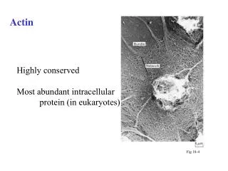
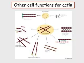
Other cell functions for actin. Actin is the most common protein in the cytoplasm. . Actin polymerization requires MgATP. Actin filaments are called microfilaments. There are over 60 actin binding proteins. Functions of Actin Filaments.

E N D
Actin is the most common protein in the cytoplasm. Actin polymerization requires MgATP. Actin filaments are called microfilaments. There are over 60 actin binding proteins. The Cytoskeleton
Functions of Actin Filaments Actin filaments are concentrated beneath the plasma membrane (cell cortex) and give the cell mechanical strength. Assembly of actin filaments can determine cell shape and cause cell movement. Association of actin filaments with myosin can form contractile structures.
Focal Adhesions Integrins anchor the outside cell surface to the extra cellular matrix by binding to proteins like fibronectin and collagen. Fibronectin----> “molecular flypaper”
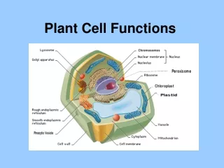
The Cytoskeleton • The cytoskeleton is an intricate network of protein filaments that extends throughout the cytoplasm. • A skin cell (fibroblast) stained with Coomassie blue.
Functions of the Cytoskeleton • Structural support for the cytoplasm • Causes changes in cell shape • Facilitates cell division • Causes cell movements • Causes muscle contraction • Controls location of organelles • Provides transport between organelles
Intermediate Filaments • Intermediate filaments enable cells to withstand mechanical stress when cells are stretched. • They can span the entire cytoplasm and are anchored to the plasma membrane.
Five Major Classes • Keratin • found in epithelial cells such as the gut and skin. Specialized keratins form hair, feathers, and claws. • Vimentin • mesodermal origin;found in connective tissue and neurological cells • Desmin • muscle cells • Glial • glial cells • Neurofilament • neurons
Microtubules http://www.reed.edu/~rsavage/microtubules.html
3-D microtubules http://www.is.kochi-u.ac.jp/Bio/cellbio/mt3d.html
Microtubules serve as "tracks" along which cells can transport organelles and vesicles. Chromosomes are pulled apart during mitosis by microtubules. Cilia and flagella are composed of microtubules. Mitotic spindle http://www.circs.neu.edu/external/Frank.Gibbons/spindle.html
Microtubules are long hollow cylinders made of the protein tubulin.
Microtubules are built from subunits of alpha and beta tubulin. • They are maintained by a balance of assembly and disassembly. • GTP (favors polymerization) or GDP
Microtubule-Associated Proteins (MAPs) bind to tubulin high molecular weight proteins, such as MAP1, MAP2 tau proteins
Capping Proteins • Capping proteins bind to the ends of microtubules and provide stabilization by protection from depolymerization
Microtubules are conveyer belts inside the cells. They move vesicles, granules, organelles like mitochondria, and chromosomes.
Motor Proteins • Motor proteins bind to microtubules and move by cycles of conformational changes using energy from ATP. • One end of the protein can bind to specific cellular components.
Microtubules provide an organizational structure in an interphase cell and separate chromosomes in a dividing cell. • Centrosome and spindle fibers
The centrosome provides nucleating sites from which microtubules can grow.
Cilia and Flagella • Cilia and flagella are composed of microtubules stabilized by association with other proteins. • Cilia move fluid over the surface of a cell. • Flagella propel single cells through fluid • Both structures use the energy of ATP to cause movement.
Ciliated cell • Microtubules can form permanent structures called cilia and flagella that provide movement to eukaryotic cells.
Adrenergic Stimulation of the Heart Figure source: http://iris.nyit.edu/~jstrauss/page2.html
Epinephrine (an endocrine secretion) and norepinephrine (a neurotransmitter) bind to adrenergic receptors. There are multiple forms of these receptors, but the myocardium is rich in G-protein linked ß-type receptors.
E Upon epinephrine or norepinephrine binding, the receptor activates the GTPase. The a subunit of the GTPase stimulates adenylate cyclase (AdC), which converts ATP into cAMP.
As the concentration of cAMP increases, a protein kinase is activated (PK-A). This phosphorylates two targets.
The calcium channels on the cell membrane become phosphorylated, increasing calcium flow into the cell. PKA also phosphorylates an inhibitory protein called phospholambam. Normally, this protein keeps the calcium ATPase transporter (calcium pump) in a low activity state. When PL is phosphorylated, it is no longer inhibitory which allows the calcium pump to operate maximally. The result is that with each beat, more calcium flows into the heart and is then removed more quickly. This translates into larger and quicker contractions.