Download
1 / 54
570 likes | 819 Views
Cell Discovery,Cells, and Cell Functions. Section Outline. Section 7-1. 7–1 Life Is Cellular A. The Discovery of the Cell 1.Early Scientist 2.The Cell Theory B. Prokaryotes and Eukaryotes 1. Prokaryotes 2. Eukaryotes C. Comparing Plant and Animal Cells.

E N D
Section Outline Section 7-1 • 7–1 Life Is Cellular A. The Discovery of the Cell 1.Early Scientist 2.The Cell Theory B. Prokaryotes and Eukaryotes 1. Prokaryotes 2. Eukaryotes C. Comparing Plant and Animal Cells
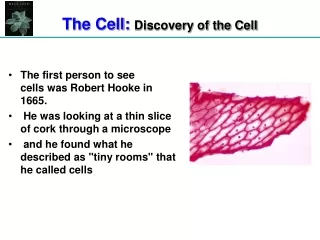
(1642) Anton von Leewenhoek invents the first microscope. • Leewenhoek became the FIRST person to OBSERVE and DESCRIBE MICROSCOPIC ORGANISMS and LIVING CELLS • (1665) Robert Hooke used a microscope to examine a thin slice of cork and described it as consisting of "a great many little boxes". Hooke named them after the latin cella-meaning “small containers” • (1838) Matthias Schleiden concluded that all PLANTS "ARE COMPOSED OF CELLS“ recognized the cell wall in protecting contents of plant cells. • ·(1838) Theodor Schwann observed cytoplasm and nucleus as vital components of animal cells • · (1855) Rudolph Virchow observed cell division (mitosis) and concluded that all cells come from preexisting cells The Discovery of Cells
The Cell Theory: ·All living things are made of cells · The cell is the smallest unit of life · All cells come from pre-existing cells · All cells exhibit the characteristics of life and have basic components (organelles) that allow them to carry out life processes and the functioning of living organisms.
Cell Theory Today Advancements in microscopic techniques, the invention of the electron microscope, and other techniques led to the continued expansion of our knowledge about the cell and its contents. Two generalizations about cells and life: • The individual cells of your organs are just as “alive” as you are, even though they cannot live independently. • The characteristics and needs of an organisms are the characteristics and needs of the cells that make up the organism.
Are All Cells Alike? Interest Grabber Section 7-1 • All living things are made up of cells, however not all cell are alike. • Some organisms are composed of only one cell (unicellular). Other organisms are made up of many cells (multicellular). • Most of the activities of a cell (repair, reproduction, etc.) are carried out via the production of proteins. • All of the organelles work together in the assemble and production of proteins. Cells are in essence “protein assemblylines”
BASIC PARTS OF A CELL Within an organism, cells differ in Size, Shape, and Organization but are similar in their DNA. • The Shape of a Cell is related to it’s Function • All Cells, regardless of function, share three basic features -all cells have an outer boundary (cell membrane) -an interior substance known as cytoplasm -and all have a genetic material(DNA).
Your Body contains at least 200 Different Cell Types. Every cell of your body has identical DNA
Two Basic Cell Types: Prokaryotic Small, bacterial cell type Lack membrane-bound nucleus (naked DNA) Lack membrane bound “organelles” with the exception of RIBOSOMES. Eukaryotic Larger, typical Plant and Animal cells Have a distinct nucleus with a membrane Contain “tiny organ-like” structures known as organelles
Similarities and Differences of Cells Prokaryotes Eukaryotes Cell membrane Cytoplasm Contain DNA Ribosomes Nucleus No nucleus Lack most organelles Organelles Plants, Animals, Fungi, Protists Bacteria Kingdoms Eubacteria and Archaebacteria
Cell membrane Cytoplasm Cell membrane Cytoplasm Prokaryotic and Eukaryotic Cells Section 7-1 Prokaryotic Cell 1000X Eukaryotic Cell Nucleus Organelles 200x
Eukaryotic Cells • Eukaryotic Cells contain a variety of structures called Organelles • Organelles PERFORM SPECIFIC FUNCTIONS FOR THE CELL. • Tissues and organs within organisms have cells that are specialized for a specific function. • Cells may have different amounts of certain organelles based on their specialized function. Examples: Muscle cells have large amounts of mitochondria White Blood Cells (Wbc’s) have large amounts of golgi and lysosomes Pancreatic cells have large amounts of Endoplasmic Reticulum
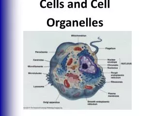
Ribosome (attached) Ribosome (free) Nucleolus Nucleus Cell Membrane Nuclear envelope Mitochondrion Smooth endoplasmic reticulum Rough endoplasmic reticulum Centrioles Golgi apparatus Figure 7-5 Plant and Animal Cells Major Organelles of Eukaryotic Cells Section 7-2 Animal Cell
Smooth endoplasmic reticulum Vacuole Ribosome (free) Chloroplast Ribosome (attached) Cell Membrane Nuclear envelope Cell wall Nucleolus Golgi apparatus Nucleus Mitochondrion Rough endoplasmic reticulum Figure 7-5 Plant and Animal Cells Major Organelles of Eukaryotic Cells Section 7-2 Plant Cell
Plasma membrane/Cell Membrane Function • Separates the cells internal fluid environment from the external fluid environment (maintains homeostasis) controlling what Enter AND Leaves the cell. • This "Selectively Permeable" Membrane regulates what passes into and out of the cell. Structure Phospholipid • TWO thin layers of Phospholipids • The TWO Phospholipid LAYERS are called a – • Phospholipid Bilayer
The Nucleus • Largest, most obvious, and darkest structure in Eukaryotic cells. • Often called the “Command Center” of the cell because it governs activities of the cell • Surrounded by a double layered membrane (nuclear envelope). • Nuclear Envelope has many small holes through which chemical messages from the nucleus can pass. • Contains DNA (Dioxyribo nucleic acid) that exists as long strands (chromatin) Most nuclei contain at least one Nucleolus a dark spot rich in RNA (ribonucleic acid). The Nucleolus- makes ribosomes which in turn build proteins During division the chromatin coils and condenses into packages known as chromosomes. Encoded information “blueprints” for building proteins is found on “genes”within the DNA .
Cytoplasm • Includes the Cytosoland the Cytoskeleton as well as the Organelles • Cytosol- “jelly-like” protein rich fluid inside the cell made up of carbohydrates, water, ions, inclusions (nonliving cell products such as fat globules). • Suspended in the cytosol are tiny Organelles (structures vital to the life of the cell) • The cytoskeleton is made up of microfilaments and microtubules that act as a scaffolding to maintain shape and provide support and help move organelles. Microtubules are hollow and are the largest strands of the cytoskeleton. Microfilaments are NOT hollow and resemble twisted rope. Microfilaments help maintain cell shape and hold organelles in place and also assist in the movement of cells.
Centrioles play a major role in cell division (mitosis) move chromosomes during mitosis bundles of microfiliments and microtubules only found in animal cells Structures for Cell Movement Flagella Cilia SHORT HAIR-LIKE PROJECTIONS that extend from the surface of the cell membrane. LONG WHIPLIKE PROJECTIONS that extend from the surface of the cell
Mitochondria The “powerhouse of the cell” Mitochondria are the sites where chemical reactions known as (Cellular Respiration) occur. Cellular Respiration transfers energy from Carbohydrates into ATP (an energy molecule used by cells) Adenosine Tri Phosphate-is the main energy currency in cells and is produced during Cellular Respiration. The structure is a large, kidney shaped, and consists double membrane. Contains long folds “cristae” for increased surface area. Also have their own DNA
Chloroplast • Specialized organelle found only in plant cells. • Often green in color due to the presence of a light trapping pigment (chlorophyll). • Sites of Photosynthesis: • Photosynthesis- the process that converts CO2 and H2O into carbohydrates using solar (light) energy • The reaction of photosynthesis take place in two stages: the light dependent stage (photophosphorylation ) and the light independent (carbon fixation/Calvin cycle) These reaction take place in membranes (grana) and the fluid filled spaces (stroma) of the thylakoid membrane system
Ribosomes • Most numerous organelle • Small spherical structures that may be “free” in the cytoplasm or attached to the membranes of the organelle known as the Endoplasmic Reticulum • Ribosomes are the site of Protein Synthesis • Often called the “protein factories of the cell” • take encoded mRna and translate the code into amino acid chains (polypeptide chains)
Endoplasmic Reticulum (EN-doh-PLAZ-mik ri-TIK-yuh-luhm) • The ER is a system of membranous tubules and sacs. • The ER functions Primarily as an Intracellular Highway, a path along which molecules move from one part of the cell to another. • ER is an extensive network of membranes that connect the Nuclear Envelope to the Cell Membrane. • The amount of ER inside a cell fluctuates, depending on the Cell's Activity. • Also helps remove Poisons, waste, and other toxic chemicals. • Transports materials through the cell.
Can be ROUGH OR SMOOTH. ROUGH ER is studded with RIBOSOMES and processes PROTEINS to be exported from the cell. SMOOTH ERIS NOT Covered with RIBOSOMES and processes LIPIDS and CARBOHYDRATES. The Smooth ER is involved in the synthesis of steroids in gland cells, the regulation of calcium levels in muscle cells, and the breakdown of toxic substances by liver cells.
Golgi apparatus • the processing, packaging and secretion organelle of the cell. • Consists of stacks of “Sac-like membranes” called Cisternae • Works closely with the ER to package proteins, antibodies, and enzymes. • Golgi apparatus is also the site of producing vesicles called Lysosomes
Lysosome • Known as “cellular garbage disposals or trash collectors” • dispose of bacteria, foreign material, and older cell components • Lysosomes are vesicules that “bud’(break-off) from the Golgi and contain digestive bags of enzymes often associated with Vacuoles • Lysosomes are the Site of Food Digestion in the Cell. They can break down large molecules such as proteins, nucleic acids, carbohydrates, and phospholipids. • Lysosomes play a very important in maintaining an organism's health by destroying cells no longer functioning properly. • Wbc’s -loaded with lysosomes
Cell Wall Chloroplasts Venn Diagrams Comparing the Plant and Animal Cells Section 7-2 Animal Cells Plant Cells Cell membrane Ribosomes Nucleus Endoplasmic reticulum Golgi apparatus Lysosomes Vacuoles Mitochondria Cytoskeleton Size and shape (large and cubic) Smaller and spherical Centrioles Motility- ability to move
Section Outline Section 7-3 • 7–3 Cell Boundaries A.Functions of the Cell Membrane B. Structure of the Cell Membrane C. Diffusion Through Cell Boundaries 1. Measuring Concentration 2. Diffusion D. Osmosis 1. How Osmosis Works 2. Osmotic Pressure E. Facilitated Diffusion F. Active Transport 1. Molecular Transport 2. Endocytosis and Exocytosis
Outside of cell Carbohydrate chains Proteins Cell membrane Inside of cell (cytoplasm) Protein channel Lipid bilayer A. Structure of the Plasma Membrane: The Cell consist of TWO thin layers of LIPIDS that separate the cell’s contents from the world around it. The two layers of PHOSPHOLIPIDS together are called the Phospholipids Bi-layer.
FLUID MOSAIC MODEL OF CELL MEMBRANES • · Membranes are Fluid (meaning they move). They are not “fixed” or rigid structures. • The Lipids and Proteins of a cell are always in motion. The phospholipids are able to drift across the membrane, changing places with neighboring phopholilids • Saturated fatty acids (solids)- more rigid cells (bone, plant tissue) • Unsaturated fatty acids (oils)- more fluid cells (muscles, skin, blood cells) • Proteins in and on the membrane form PATTERNS, or “MOSAICS”. • · Mosaic meaning it is made of patterns of Channel Proteins, Glycolipids, and Cholesterol molecules contribute to the fluidity or rigidity of the cell • Scientists view the plasma membrane as a “Fluid - Mosaic Model”
Outside of cell Carbohydrate chains Proteins Cell membrane Inside of cell (cytoplasm) Protein channel Lipid bilayer Membrane Proteins A Variety of PROTEIN MOLECULES are EMBEDDED in the Lipid Bilayer Integral Proteins that serve as “Doorways” and “Windows” to the cells interior often have carbohydrates attached to serve as identification badges that allow cells to recognize each other and may act as Site where viruses or chemical messengers such as hormones can attach.
Homeostasis and the Plasma Membrane • 1. Functions of the Plasma (cell) Membrane • ·Separates the cells internal fluid environment from the external fluid environment (maintains homeostasis). It controls what Enters AND Leaves the cell. • · Transports water, ions, gases, and nutrients based on size and composition • Protects the internal organelles receives communication from other cells
Homeostasis and the Plasma Membrane 2. Selective permeability • The cell membrane allows the passage of some molecules but is impermeable to others. • Movement can occur actively (spending energy) or passively (without spending energy). • Direction of movement depends on the types and concentrations of dissolved solutes both in and outside the cell.
Homeostasis and the Plasma Membrane 3. Concentration gradient • The concentration of a solution is the amount of solute (dissolved substance) in a given amount of solution (usually water). • A Concentration Gradient is the differences in concentration across a membrane • In a cell, dissolved solutes move from High concentrations to Low concentrations- passive transport • Movement against the concentration gradient (lowto high) requires energy- active transport
Molecule to be carried Energy Molecule being carried Active transport • -movement against the diffusion concentration gradient • -movement from area of low concentration to area of high concentration. • -requires the expenditure of energy (uses ATP from mitochondria to “pump” in ions and molecules
4.Types of Diffusion Through the Cell Membrane 7-12 The Structure of the Cell Membrane Section 7-3 • Small molecules like Oxygen and Carbon Dioxide enter passively (without energy expenditure) • Types of diffusion: • Simple Diffussion- movement from an area of high (greater) concentration to an area of low (lesser) concentration • Large particles like glucose, protein, and certain ions enter through protein channels- facilitated diffusion (enzyme or protein regulated but still passive) • Osmosis- diffusion of water in response to differences in solute concentrations across a membrane.
Facilitated Diffusion- movement of large molecules ·Large particles like glucose, protein, and certain ions enter through protein channels- facilitated diffusion (enzyme or protein regulated but still passive) Glucose molecules High Concentration Cell Membrane Low Concentration Protein channel
Figure 7-15 Osmosis Section 7-3 When dissolved molecules (solutes) are too large to pass through the cell membrane, cells respond by passively moving water into and out of the cell by Osmosis
5. When does diffusion stop? When the concentration of the dissolved solute on one side of the membrane is = to the concentration of the dissolved solute on the opposite side of the membrane, the system has reached Dynamic Equilibrium Recap: Movement in diffusion: high concentrationlow concentration until all solutes are evenly distributed (=)
Types of Solutions • Isotonic solution- the concentration of dissolved solutes (sugar) and solvent (water) will be the same on both sides of the membrane. • Hypertonic solution- the concentration of dissolved solute (sugar) is greater in the solution than the concentration of the solvent (water). • Hypotonic solution- the concentration of dissolved solute (sugar) is less in the solution than the concentration of the solvent (water).
Bulk transport a) Endocytosis - Membrane surrounds and engulfs by forming a sealed vacuole. Two types of endocytosis: a) Phagocytosis- “cell eating” b) Pinocytosis- “cell drinking” b) Exocytosis -discharge of wastes from the cells interior.
Ameba proteus- eats bacteria and other unicellular organisms by phagocytosis White blood cells (phagocytes) eat and destroy bacteria