Download
1 / 6

E N D
Hairy cell leukemia, blastic • 54-year-old male, admitted to the emergency room in May 2008 with acute pain in the upper-left quadrant and constitutional syndrome. The examination revealed a ruptured spleen and lymphadenopathies involving cervical, retroperitoneal and mesenteric lymph nodes. No autoimmune disorders were reported. • Laboratory studies showed leukocytosis (19.81 x 10⁹/L) with 5.48 x 10⁹/L lymphocytes, 125 x 109/L platelets, 10.7 g/dL hemoglobin, 2,130 U/L LDH, 1,470/493/21 IgG/A/M, 32 g/L albumin.
Hairy cell leukemia, blastic • PB morphology indicated the presence of 16% large cells, each with a round-to-oval nucleus, no convolutions, an eccentric disposition and an inconspicuous central nucleolus. Some cells exhibited a blastic morphology and others featured villous (HCL-like) projections. The nucleus/cytoplasm ratio was relatively high. TRAP was weakly positive. • FCM was positive for CD11c, CD20, CD25, CD103 and CD123, with kappa restriction. • Splenectomy was performed (1,450 g, 26 x 15 x 8 cm) and the patient received adjuvant chemotherapy with CHOP. • Postoperatively he developed cholostasis with increased hepatic enzymes, which led to septic shock, multiple organ failure and, finally, the demise of the patient.
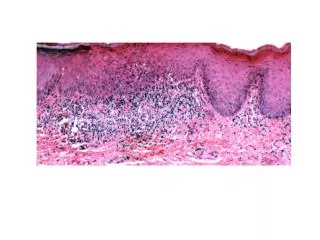
Hairy cell leukemia, blastic • Bone-marrow infiltration had an interstitial and diffuse pattern. • Spleen histology revealed extensive infiltration of the red pulp and effacement of the white pulp. Neoplastic cells were of medium and large size, with abundant cytoplasm. Their nuclei were large and had irregular contours and dispersed chromatin with inconspicuous nucleoli. • The immunohistochemical study revealed expression of Annexin A1, bcl2, CD20, DBA44, and IgG, and an absence of bcl6, CD10, CD23, CD5, Cyclin D1, IgD and p53. Ki-67 was relatively high. • Karyotypic studies revealed numerical and structural abnormalities of a cellular clone with 48 chromosomes, and the presence of rearrangements involving low point breakage at 3q27, 8q24 and 18q21, where the genes BCL6, MYC and BCL2, respectively, were located.
Hairy cell leukemia, blastic • HCL – aggressive variant? • HCL – blastic variant?