Chapter 8 Antigen Processing and Presentation
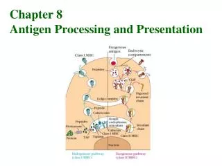
Chapter 8 Antigen Processing and Presentation. 本章大綱 : 1. Self-MHC Restriction of T Cells 2. Role of Ag-Presenting Cells 3. Evidence for Two Processing and Presentation Pathways 4. Endogenous Ags: The Cytosolic Pathway 5. Exogenous Ags: The Endocytic Pathway

Chapter 8 Antigen Processing and Presentation
E N D
Presentation Transcript
Chapter 8 Antigen Processing and Presentation
本章大綱: 1. Self-MHC Restriction of T Cells 2. Role of Ag-Presenting Cells 3. Evidence for Two Processing and Presentation Pathways 4. Endogenous Ags: The Cytosolic Pathway 5. Exogenous Ags: The Endocytic Pathway 6. Presentation of Nonpeptide Bacterial Ags
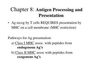
Self-MHC Restriction: T cells can only recognize Ag when it is presented with a self-MHC molecule on the membrane of an APC.
Self-MHC restriction of TH cells APC T cell class II MHC restriction
Self-MHC restriction of TC cells class I MHC restriction
When a primary Ab response and cell-mediated response were induced by a protein in its native form, a secondary Ab response could be induced only by native Ag, whereas a secondary cell- mediated response could be induced by either the native or the denatured Ag.
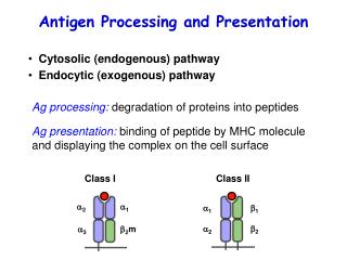
Necessity of Ag Processing (para- Formaldehyde) Ag processing involves the digestion of the protein into peptides. (glutaraldehyde)
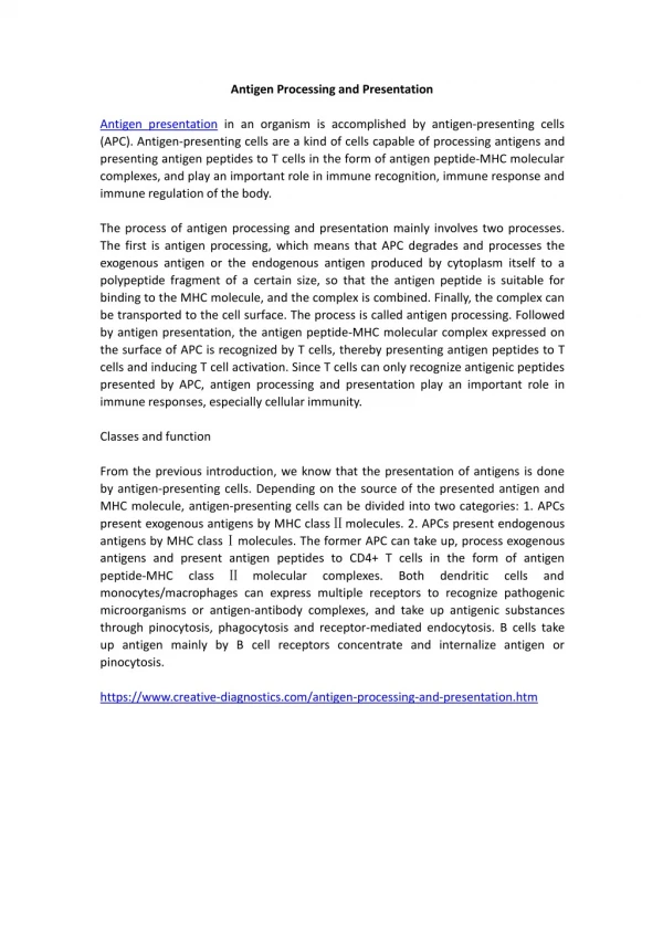
Since all cells expressing either class I or class II MHC molecules can present peptides to T cells, strictly speaking they all could be designated as Ag-presenting cells (APC). However,………………..
Ag-presenting cells (APC): Cells that display peptides associated with class II MHC molecules to CD4+ TH cells are called APC. Target cells: Cells that display peptides associated with class I MHC molecules to CD8+ TC cells are referred to as target cells.
Dendritic cells: 1. constitutively express a high level of class II MHC molecules and costimulatory activity 2. most effective in Ag presentation
Because nearly all nucleated cells express class I MHC molecules, virtually any nucleated cell is able to function as target cell presenting endogenous Ags to CD8+ T cells. Most often, target cells are cells that have been infected by a virus or some other intracellular microorganisms.
emetine: inhibits viral protein synthesis chloroquine: blocks the endocytic processing pathway
Cytosolic proteolytic system for degradation of intracellular proteins
Transporters Associated with Ag Presentation (TAP)
Both TAP1 and TAP2 belong to the family of ATP-binding cassette (ABC) proteins found in the membranes of many cells, including bacteria. These proteins mediate ATP-dependent transport of amino acids, sugars, ions, and peptides.
Generation of antigenic peptides in the cytosolic pathway Proteosome subunits (LMP2, LMP7, and LMP10) favor the production of peptides that binds to class I MHC molecules. b2M
LMP and TAP genes in Mouse H-2 LMP and TAP genes in mouse H-2 Class I Nonclassical Class IIClass IIIClass I Nonclassical LMP: 1. LMP 2 and LMP7 are encoded within MHC and induced by interferon g. 2. LMP10 is not encoded within MHC and also induced by interferon g. 3. LMPs preferentially generate peptides that bind to class I MHC molecules. Such proteosomes show increased hydrolysis of peptide bonds that follow basic and/or hydrophobic residues, the preferred anchor residues for class I. 4. Polymorphic TAP: 1. has the highest affinity for peptides containing 8-13 amino acids. 2. favors peptides with basic and/or hydrophobic C-terminal a. a. 3. Thus, TAP is uniquely designed to transport peptides that will interact with class I MHC molecules. 4. polymorphic
Assembly and stabilization of class I MHC molecules Molecular chaperones: calnexin: a redident membrane protein of the ER calreticulin tapasin (TAP-associated protein) : brings the TAP into proximity with the class I molecules.
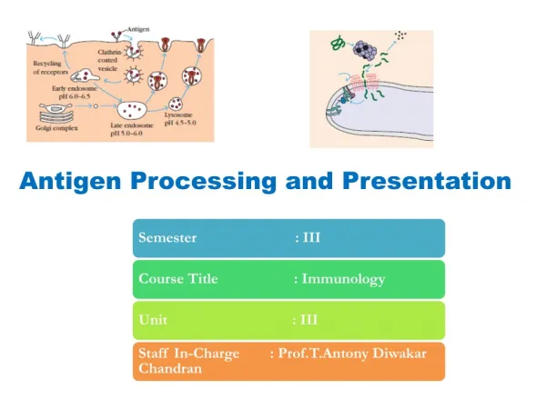
Generation of antigenic peptides in the endocytic pathway • 40 acid- dependent • hydrolases in • lysosomes: • 1. proteases • 2. nucleases • 3. glycosidases • 4. lipases • 5. phospholipases • 6. phosphatases
Assembly of class II MHC molecules Facilitate the loading of peptides into class II a negative regulator, inhibiting the reaction between CLIP and HLA-DM CLIP :class II-associated invariant chain peptide
Functions of invariant (Ii) chain: • Prevention of the binding of endogenous peptides to class II molecules. • Proper folding of the class II a and b chains • Exit of class II from the RER • Routing of class II to the endocytic processing pathway from the trans-Golgi.
HLA-DM and HLA-DO genes in Human HLA Class II Nonclassical Class IIClass IIIClass I Nonclassical Nonclassical class II genes : DM : 1. encode class II-like molecules that facilitate the loading of antigenic peptides into the class II MHC molecules 2. expressed within the endosomal compartments 3. not polymorphic DO : 1. limited expression in thymus and in mature B cells 2. a regulator of class II antigen processing. 3. Not polymorphic
3-D structure of the binding groove of class II MHC molecules Red : DR4 + collagen II peptide Yellow : DR1 + influenza hemagglutinin peptide Blue : DR3 + CLIP
Presentation of nonpeptide Ags by class I-like CD1 molecules