Download

1 / 15
240 likes | 834 Views
Neuromuscular Junction. Figure 9.7 (a-c). Skeletal Muscle. Figure 9.2 (a). Myofibrils. Figure 9.3 (b). Sarcomeres. Figure 9.3 (c). Myofilaments: Banding Pattern. Figure 9.3 (c, d). Ultrastructure of Myofilaments: Thick Filaments. Figure 9.4 (a)(b).

E N D
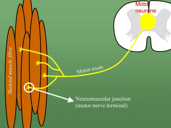
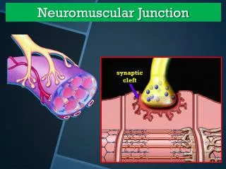
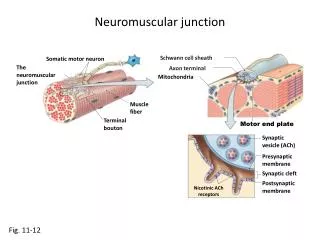
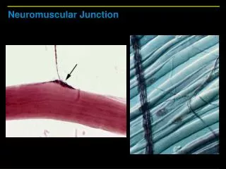
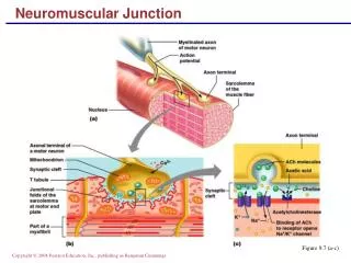
Neuromuscular Junction Figure 9.7 (a-c)
Skeletal Muscle Figure 9.2 (a)
Myofibrils Figure 9.3 (b)
Sarcomeres Figure 9.3 (c)
Myofilaments: Banding Pattern Figure 9.3 (c, d)
Ultrastructure of Myofilaments: Thick Filaments Figure 9.4 (a)(b)
Ultrastructure of Myofilaments: Thin Filaments Figure 9.4 (c)
Arrangement of the Filaments in a Sarcomere • Longitudinal section within one sarcomere Figure 9.4 (d)
Sarcoplasmic Reticulum (SR) Figure 9.5
Role of Ionic Calcium (Ca2+) in the Contraction Mechanism • At low intracellular Ca2+ concentration: • Tropomyosin blocks the binding sites on actin • Myosin cross bridges cannot attach to binding sites on actin • The relaxed state of the muscle is enforced Figure 9.10 (a)
Role of Ionic Calcium (Ca2+) in the Contraction Mechanism • At higher intracellular Ca2+ concentrations: • Additional calcium binds to troponin (inactive troponin binds two Ca2+) • Calcium-activated troponin binds an additional two Ca2+ at a separate regulatory site Figure 9.10 (b)
Role of Ionic Calcium (Ca2+) in the Contraction Mechanism • Calcium-activated troponin undergoes a conformational change • This change moves tropomyosin away from actin’s binding sites Figure 9.10 (c)
Role of Ionic Calcium (Ca2+) in the Contraction Mechanism • Myosin head can now bind and cycle • This permits contraction (sliding of the thin filaments by the myosin cross bridges) to begin Figure 9.10 (d)
Sequential Events of Contraction Myosin head (high-energy configuration) Myosin cross bridge attaches to the actin myofilament 1 Thin filament ADP and Pi (inorganic phosphate) released Thick filament Working stroke—the myosin head pivots and bends as it pulls on the actin filament, sliding it toward the M line As ATP is split into ADP and Pi, cocking of the myosin head occurs 2 4 Myosin head (low-energy configuration) As new ATP attaches to the myosin head, the cross bridge detaches 3 Figure 9.11
Motor Unit: The Nerve-Muscle Functional Unit Figure 9.12 (a)