Download
1 / 25
250 likes | 363 Views
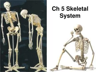
Ch. 6 The Skeletal System. Introduction. Skeletal System Bones Structures that make up the joints Tissues Bone Cartilage Fibrous conn . tissue – forms ligaments that connect bone to bone. Functions of the Skeleton. Supports the body Protects some internal organs

E N D
Introduction • Skeletal System • Bones • Structures that make up the joints • Tissues • Bone • Cartilage • Fibrous conn. tissue – forms ligaments that connect bone to bone
Functions of the Skeleton • Supports the body • Protects some internal organs • Contains & protects red bone marrow • Stores excess calcium
Types of Bone Tissue • Bone cells – osteocytes • Matrix – calcium salts, collagen • In normal circumstances, calcium removed = calcium deposited • Regulated by osteocytes • Fig. 6-1 shows 2 types of bone tissue • Compact (ground) • Cancellous (spongy)
Types of Bone Tissue (cont.) • Compact • Made of osteons (haversian systems) • Microscopic cylinders of bone matrix with osteocytes in concentric rings around central haversian canals • Microscopic channels (canaliculi) connect osteocytes with blood vessels and other osteocytes
Types of Bone Tissue (cont.) • Spongy • No haversian system • Contains osteocytes, matrix, blood vessels • Cavities often contain red bone marrow, which produce RBCs, platelets, WBCs
Classification of Bones • Long • Short • Bones of wrists, ankles • Flat • Ribs, shoulder blades, hip bones, cranial bones • Irregular • Vertebrae, facial bones
Classification of Bones (cont.) • Long Bones • Bones of arms, legs, hands, feet • Shaft (diaphysis) • Made of compact bone; hollow, forming a canal within the shaft • Marrow canal (medullary cavity) contains yellow bone marrow (adipose tissue) • Ends (epiphyses) (Fig. 6-1) • Made of spongy bone covered with thin layer of compact bone • Red bone marrow replaced by yellow in adult bones
Classification of Bones (cont.) • Short, flat, irregular bones made of spongy bone covered with thin layer of compact bone • Red bone marrow within spongy bone • Joint surfaces covered with articular cartilage • Periosteum covers remainder of bone • Fibrous conn. tissue membrane; its collagen fibers merge with tendons & ligaments attached to bone • Anchors aforementioned structures and contains blood vessels & osteoblasts
Embryonic Growth of Bone • Skeleton is first made of cartilage & fibrous connective tissue; gradually replaced by bone • Bone matrix produced by osteoblasts • Process called ossification • Baby has fibrous connective tissue between skull bones (fontanels); Fig. 6-2 • Permit compression during birth, growth of brain after birth • These “soft spots” are ossified by the age of 2
Embryonic Growth of Bone (cont.) • Rest of embryonic skeleton • First made of cartilage • ossification begins in 3rd month of gestation in long bones • Osteoblasts produce bone matrix in center of diaphyses of long bones and in center of short, flat, irregular bones (Fig. 6-3)
Embryonic Growth of Bone (cont.) • Long bones develop centers of ossification in epiphyses • Growth occurs in epiphyseal discs (cartilage) at jxn of diaphysis with epiphysis • Bone grows in length as more cartilage is produced on epiphysis side (Fig. 6-3) • On diaphysis side, osteoblasts produce bone matrix to replace cartilage • B/w ages 16-25, all cartilage of epiphyseal disc is replaced by bone; bone lengthening stops
Embryonic Growth of Bone (cont.) • Osteoclasts dissolve & reabsorb minerals of bone matrix (resorption) • Very active in embryonic long bones • Reabsorb bone matrix in center of diaphysis to form marrow canal • Blood vessels grow into marrow canals of embryonic long bones, establishing red bone marrow • After birth, red marrow replaced by yellow • Red marrow remains in spongy bone of short, flat, irregular bones
Factors that Affect Bone Growth & Maintenance • Heredity – polygenic inheritance • Nutrition – Ca, P, protein, vitamin A, C, D • Hormones – growth hormone, thyroxine, parathyroid hormone, insulin, estrogen, testosterone (Table 6-1) • Exercise or “stress” – bearing weight • As simple as everyday walking
The Skeleton • 2 divisions • Axial – forms body’s axis • Skull, vertebral column, rib cage • Appendicular – supports appendages or limbs • Bones of arms, legs, shoulder, pelvic girdles • 206 bones in body (Fig. 6-4)
The Skeleton • Study Fig. 6-4 thru 6-16 • Study Tables 6-2 thru 6-5
Joints - Articulations • Joint – where 2 bones meet or articulate • See Table 6-5 & Fig. 6-15 • Classification of joints • Synarthrosis – immovable joint (suture b/w 2 cranial bones) • Amphiarthrosis – slightly movable joint (symphysis joint b/w adjacent vertebrae) • Diarthrosis – freely movable joint; largest category • Ball & socket • Pivot • Hinge, etc.
Synovial Joints • All diarthroses are synovial joints (Fig. 6-16) • On the joint surface of each bone is articular cartilage, providing a smooth surface • Joint capsule of fibrous connective tissue encloses joint in a strong sheath • Synovial membrane lines joint capsule, secreting synovial fluid into joint cavity • Fluid is thick & slippery, preventing friction
Synovial Joints • Many have bursae • Small sacs of synovial fluid b/w joint & tendons that cross over the joint • Permit tendons to slide easily as bones are moved • Bursitis • Excessive use of a joint, causing inflammation & pain
Aging & The Skeletal System • Bone tissue loses Ca • Bone matrix thins • Bones become brittle • Fractures are more likely to occur • Erosion of articular cartilage • Knees, fingers • What can you do? • Exercise • Diet high in Ca, Vitamin D