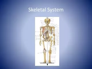
Ch 5f: The Skeletal System
260 likes | 651 Views
Ch 5f: The Skeletal System. Structural Classification of Joints. Fibrous joints Generally immovable Cartilaginous joints Immovable or slightly moveable = amphiarthroses Synovial joints Freely moveable. Fibrous Joints. Bones united by fibrous tissue Examples Sutures of skull Syndesmoses

Ch 5f: The Skeletal System
E N D
Presentation Transcript
Structural Classification of Joints • Fibrous joints • Generally immovable • Cartilaginous joints • Immovable or slightly moveable = amphiarthroses • Synovial joints • Freely moveable Hoban
Fibrous Joints • Bones united by fibrous tissue • Examples • Sutures of skull • Syndesmoses • Allows more movement than sutures • Example: distal end of tibia and fibula Figure 5.27a–b Hoban
Fibrous Joints Hoban
Fibrous Joint: Sutures Hoban
Cartilaginous Joints – amphiarthrotic (slightly moveable) • Bones connected by cartilage • Examples • Pubic symphysis • Intervertebral joints Figure 5.27d–e Hoban
Cartilaginous Joints Hoban
Synovial Joints • Articulating bones are separated by a joint cavity • Synovial fluid is found in the joint cavity • Joints of limbs Figure 5.24f–h Hoban
Synovial Joints Hoban
Features of Synovial Joints • Articular cartilage (hyaline cartilage) covers the ends of bones • Joint surfaces are enclosed by a fibrous articular capsule • Have a joint cavity filled with synovial fluid • Ligaments reinforce the joint Hoban
Synovial Joint: Elbow Hoban
Synovial Joint: Hip Hoban
Synovial Joint: Knee Hoban
Synovial Joint: Shoulder Hoban
Structures Associated with the Synovial Joint • Bursae – flattened fibrous sacs • Lined with synovial membranes • Filled with synovial fluid • Not actually part of the joint • Tendon sheath • Elongated bursa that wraps around a tendon Hoban
The Synovial Joint Figure 5.28 Hoban
Types of synovial joints based on shape • Shape of articulating bone determines movements • Plane • Flat surfaces • Gliding movements • Intercarpal joints of wrist Hoban
Hinge • Cylindrical end of one with trough-shaped surface of 2nd • Movement in one plane • Elbow, ankle, phalanges • Pivot • Rounded end of one bone fits into sleeve of other • Rotate around axis • Proximal radioulnar joint, axis & atlas Hoban
Figure 5.29a–c Hoban
Condyloid • Egg shape of one fits into oval concavity of 2nd • Move back & forth but not rotate • Fingers • Saddle • Convex & concave areas form saddle • thumb Hoban
Ball & socket • Sphere of one into socket of 2nd • Movement in all axes • Most freely moving • Shoulder & hip Hoban