Download
1 / 52
550 likes | 1.24k Views
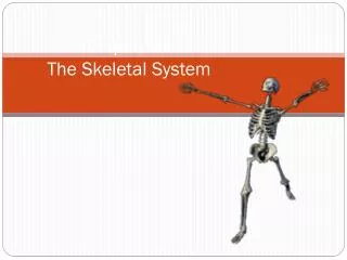
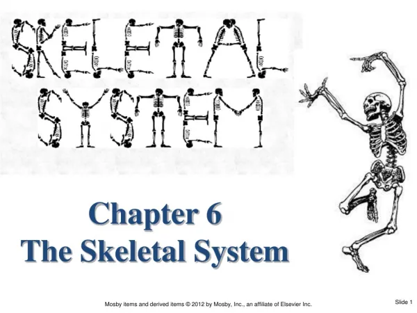
Chapter 6 The Skeletal System. I. FUNCTIONS - SKELETAL SYSTEM. Supports and gives shape to the body. I. FUNCTIONS - SKELETAL SYSTEM. Supports and gives shape to the body. Protects internal organs. I. FUNCTIONS - SKELETAL SYSTEM. Supports and gives shape to the body.

E N D
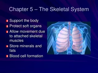
I. FUNCTIONS - SKELETAL SYSTEM Supports and gives shape to the body.
I. FUNCTIONS - SKELETAL SYSTEM Supports and gives shape to the body. Protects internal organs.
I. FUNCTIONS - SKELETAL SYSTEM Supports and gives shape to the body. Protects internal organs. Helps make movements possible when bones at moveable joints are pulled by muscles.
I. FUNCTIONS - SKELETAL SYSTEM Storescalcium — hormones regulate calcium storage:
I. FUNCTIONS - SKELETAL SYSTEM Storescalcium — hormones regulate calcium storage: calcitonin (CT) increases storage parathyroid hormone (PTH) reduces stores of calcium
I. FUNCTIONS - SKELETAL SYSTEM Storescalcium — hormones regulate calcium storage: calcitonin (CT) increases storage parathyroid hormone (PTH) reduces stores of calcium Hematopoiesis(hee-MA-toh-poy-EE-sis) —blood cell formation in red bone marrow
II. TYPES OF BONES A. Four major types, according to overall shape of the bone Long —Example: humerus (arm)
II. TYPES OF BONES A. Four major types, according to overall shape of the bone Long —Example: humerus (arm) Short —Example: carpals (wrist)
II. TYPES OF BONES A. Four major types, according to overall shape of the bone Long —Example: humerus (arm) Short —Example: carpals (wrist) Flat —Example: frontal (skull)
II. TYPES OF BONES A. Four major types, according to overall shape of the bone Long —Example: humerus (arm) Short —Example: carpals (wrist) Flat —Example: frontal (skull) Irregular —Example: vertebrae (spinal cord)
II. TYPES OF BONES A. Four major types, according to overall shape of the bone Long —Example: humerus (arm) Short —Example: carpals (wrist) Flat —Example: frontal (skull) Irregular —Example: vertebrae (spinal cord) Some also recognize a sesamoid (round) bone category — Example: patella (kneecap)
II. TYPES OF BONES B. Structure of long bones (Figure 6-1) Diaphysis (dye-AF-i-sis) or shaft —hollow tube of hard compact bone
B. Structure of long bones (Figure 6-1) Medullarycavity —hollow area inside diaphysis bone that contains yellowmarrow (inactive, fatty marrow found in adults) II. TYPES OF BONES
B. Structure of long bones (Figure 6-1) Epiphyses, (eh-PIF-i-seez) or ends of the bone —spongy bone that contains red bone marrow II. TYPES OF BONES
B. Structure of long bones (Figure 6-1) Articularcartilage —covers epiphyses and functions as a cushion II. TYPES OF BONES
B. Structure of long bones (Figure 6-1) Periosteum —strong membrane covering bone everywhere except at joint surfaces II. TYPES OF BONES
B. Structure of long bones (Figure 6-1) Endosteum —thin membrane lining medullary cavity II. TYPES OF BONES
C. Structure of flat bones Spongy bone layer sandwiched between two compact bone layers Diploe (DIP-lo-ee) — spongy bone layer of a flat bone II. TYPES OF BONES
MICROSCOPIC STRUCTURE OF BONE AND CARTILAGE Bone types (Figure 6-3) Spongy Texture results from needlelike threads of bone called trabeculae surrounded by a network of open spaces Found in epiphyses of bones Spaces contain red bone marrow Compact Structural unit is an osteon—calcified matrix arranged in multiple layers or rings called concentric lamella (Figure 6-4) Bone cells are called osteocytes and are found inside spaces called lacunae, which are connected by tiny tubes called canaliculi
MICROSCOPIC STRUCTURE OF BONE AND CARTILAGE (cont.) Cartilage (Figure 6-5) Cell type called chondrocyte Matrix is gel-like and lacks blood vessels
BONE FORMATION AND GROWTH Early bone development (before birth) consists of cartilage and fibrous structures Osteoblasts form new bone, and osteoclasts reabsorb bone; osteocytes are inactive osteoblasts (Figure 6-6) Cartilage models gradually replaced by calcified bone matrix—process called endochondral ossification (Figures 6-7 and 6-8)
DIVISIONS OF SKELETON Skeleton composed of the following divisions and subdivisions: Axial skeleton Skull Spine (vertebral column) Thorax Hyoid bone Appendicular skeleton Upper extremities, including shoulder (pectoral) girdle Lower extremities, including hip (pelvic) girdle Location and description of bones—see Figures 6-9 to 6-20 and Tables 6-2 to 6-6
DIFFERENCES BETWEEN A MAN’S AND A WOMAN’S SKELETON Size—male skeleton generally larger Shape of pelvis—male pelvis deep and narrow; female pelvis broad and shallow Size of pelvic inlet—female pelvic inlet generally wider, normally large enough for baby’s head to pass through it (Figure 6-21) Pubic angle—angle between pubic bones of female generally wider
JOINT (ARTICULATIONS) Every bone except the hyoid (which anchors the tongue) connects to at least one other bone Kinds of joints (Figures 6-22 to 6-24) Synarthroses (no movement)—fibrous connective tissue grows between articulating bones; example: sutures of skull Amphiarthroses (slight movement)—cartilage connects articulating bones; example: symphysis pubis
JOINT (ARTICULATIONS) (cont.) Kinds of joints (cont.) Diarthroses (free movement)—most joints belong to this class Structure Structures of freely movable joints—joint capsule and ligaments hold adjoining bones together but permit movement at joint Articular cartilage—covers joint ends of bones and absorbs joints Synovial membrane—lines joint capsule and secretes lubricating fluid Joint cavity—space between joint ends of bones Bursa—fluid-filled pouch that absorbs shock; inflammation of bursa is called bursitis Functions of freely moveable joints—ball-and-socket, hinge, pivot, saddle, gliding, and condyloid—allow different kinds of movements determined by the structure of each joint (Table 6-7)