Download
1 / 81
850 likes | 1.14k Views
Functional Anatomy of the Basal Ganglia. Sharif Taha, Ph.D. s.taha@utah.edu Department of Neurobiology and Anatomy. Outline. Anatomy a. BG components b. Anatomical connectivity Function: Modulation through disinhibition Action Selection Neuromodulators: dopamine.

E N D
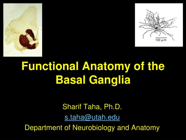
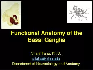
Functional Anatomy of the Basal Ganglia Sharif Taha, Ph.D. s.taha@utah.edu Department of Neurobiology and Anatomy
Outline • Anatomy a. BG components b. Anatomical connectivity • Function: Modulation through disinhibition • Action Selection • Neuromodulators: dopamine
What do the basal ganglia do? • Modulate the initiation, termination, amplitude, and selection of movement - Initiation and selection 2. Learning -Response-outcome associations - Stimulus-response associations
Basal ganglia: a modulatory cortical loop • Basal Ganglia receives robust input from the cortex - Almost all parts of cortex; excludes primary sensory cortices • Principal projection of the BG - back to cortical targets - Motor associated areas - Via ventral thalamic relay (Other targets: superior colliculus)
Overview of BG organization • Output: • Substantia nigra pars reticulata (SNr) • Internal segment of globus pallidus (GPi) • Neuromodulator: • Substantia nigra pars compacta (SNc) • Input: • Caudate and putamen (together, the striatum) • Intrinsic: • Subthalamic nucleus (STN) • External segment of globus pallidus (GPe) SNc
Striatum: Medium spiny neurons • Caudate and putamen • Medium spiny neurons • ~90% of neurons; primary projection neurons • GABAergic; inhibitory • Very little spontaneous activity; dependent on excitatory input for discharge
Up and down states • Inwardly rectifying potassium channels keep striatal neurons (very) hyperpolarized • Membrane potential shifts from hyperpolarized potentials (−80 mV) to more depolarized potentials (−50mV) • Transitions to the up state are correlated among nearby striatal neurons • Selection mechanism – requires concerted cortical activation to move to upstate Wilson 1998 Science
Striatum: Intrinsic interneurons 2 principle types • 3 GABAergic interneurons • Tonically active neurons (TANs) • Cholinergic • Large cell bodies
Globus pallidus Two segments → Internal: Principle output nucleus →External: intrinsic circuitry Neurons in both areas - high tonic firing rates GABAergic, inhibitory
Subthalamic nucleus Alone among the BG circuit elements –glutamatergic Target for deep brain stimulation (DBS)
Nigral Complex • Midbrain • Substantia nigra pars reticulata (SNpr) • GABAergic • Output of BG • Developmentally, related to Gpi • Substantia Nigra pars Compacta (SNpc) • Neuromelanin-containing cells • Dopaminergic (A9) SNc
Basal ganglia connectivity Cortical input Thalamus Cortex Subthalamic nucleus
Three organizing principles of basal ganglia connectivity Cortical input • Anatomically parallel loops with distinct function • Finer-grain topographic organization within loops • Patch/matrix Thalamus Cortex Subthalamic nucleus
Functional topography: Parallel loops w/in the BG subserve distinct functions
Functional topography: Parallel loops w/in the BG subserve distinct functions • 4 pathways: • Skeletomotor • Oculomotor channel • Association • Behavior, learning, cognition • Limbic • Addiction, emotional behavior • J.H. Martin, Neuroanatomy: Text and Atlas 2nd Ed., 1996
Topography is also maintained within loops: Somatotopy • J.H. Martin, Neuroanatomy: Text and Atlas 2nd Ed., 1996
Oculomotor topography • J.H. Martin, Neuroanatomy: Text and Atlas 2nd Ed., 1996
Patch/matrix compartments: neurochemical organization • Neurochemically distinct areas (patch, mu opioid receptor; matrix, calbindin) • Dendrites observe boundaries • Afferents/efferents are distinct • Functional roles – • Patch: limbic • Matrix: sensorimotor
Outline • Anatomy a. BG components b. Anatomical connectivity • Modulating action through disinhibition • Direct and Indirect Pathways • Action Selection • Neuromodulators • Pathology
Output nuclei maintain a high tonic level of discharge, suppressing activity in target regions
Firing under quiescent conditions (in the absence of movement)
Movement modulation occurs through disinhibition of thalamocortical target regions
What advantages does modulation through inhibition confer? • Strong tonic inhibition allows basal ganglia to serve as a master regulator – arbitrating between multiple excitatory inputs • Initiating and • Discriminating Cortical regions Saccade generator
Basal ganglia: movement modulation through disinhibition • Output nuclei of the basal ganglia are inhibitory • Output nuclei maintain a high tonic level of discharge, suppressing activity in target regions • Phasic decrease in firing rate transiently releases target regions from inhibition. • Disinhibited thalamocortical circuit discharges, promoting movement.
Outline • Anatomy a. BG components b. Anatomical connectivity • Modulating action through disinhibition • Direct and Indirect Pathways • Action Selection • Neuromodulators • Pathology
Basal firing rates in the striatum are very low,and dependent upon strong cortical excitation.
Under these conditions, striatal firing has little impact on GPi/SNr discharge
Phasic cortical excitation drives excitatory discharge in the striatum.
Activation of the direct pathway promotes action. This causes a transient inhibition of GPi/SNr firing.
Striatal neurons have low tonic firing rates; again, dependent upon strong cortical inputs
GPe neurons are similar to those in GPi; they have high tonic firing rates
Firing under quiescent conditions (in the absence of movement)
Followed by phasic excitation of the STN (through disinhibition)…
And finally, a increased rate of discharge in the output nuclei - Activation of the indirect pathway suppresses action.
Rate model & basal ganglia pathology http://www.youtube.com/watch?feature=player_detailpage&v=fCL7RWaC3RA http://www.youtube.com/watch?feature=player_detailpage&v=AvBrP4yRTRA
Indirect pathway suppresses action. Direct pathway facilitates action. How do they cooperatively regulate motor output?
Outline • Anatomy a. BG components b. Anatomical connectivity • Modulating action through disinhibition • Direct and Indirect Pathways • Action Selection • Neuromodulators • Pathology