Basal Ganglia
320 likes | 709 Views
Basal Ganglia. CD-ROM: Case II, The Shaky Carpenter Notes: Chapter 4, Basal Ganglia, p. 28-32. Case Description. Case 2: The Shaky Carperter, Case Description, frame 1-3. CNS centres that participate in the commend of skeletal muscle. Ideomotor area Premotor cortex

Basal Ganglia
E N D
Presentation Transcript
Basal Ganglia CD-ROM: Case II, The Shaky Carpenter Notes: Chapter 4, Basal Ganglia, p. 28-32
Case Description Case 2: The Shaky Carperter, Case Description, frame 1-3
CNS centres that participate in the commend of skeletal muscle • Ideomotor area • Premotor cortex • Primary motor cortex • Basal ganglia • Cerebellum • Spinal reflexes
Basal Ganglia: Basic Functions • participate in planning of high-level movement synergies, including initiation and termination of movements. Background: Frame 1
Basal Ganglia: Objectives • 1. Categorize and list the nucleic components ofbasal ganglia. • . • 2. Provide a general description of the circuitry through the nuclei*. • Illustrate theanatomical relationshipsof the nuclei • AND • their relationships to important neighbouring structures. *FYI only
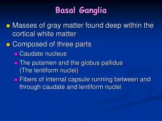
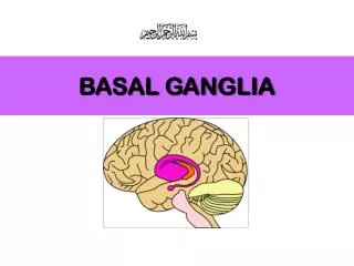
Objective 1a: Components of the Basal Ganglia • Corpus Striatum (telencephalic): • a. Striatum • i. Caudate nucleus* • ii. Putamen* • b. Pallidum • i. Globus Pallidus (External)* • ii. Globus Pallidus (Internal)* • 2. Subthalamic Nucleus (diencephalic) • 3. Substantia nigra (mesencephalic) Frame 2,3 Notes: p. 28,Section 4.1.1 and p.29, Section 4.2.1
Objective 1b: Taxonomy of Corpus Striatum Lentiform nucleus Anatomically identifiable entities Frame 2-3 Notes: p. 20, Section 4.2.1
Objective 2: Circuitry between nuclei of the Basal Ganglia (FYI only) Cerebral cortex Motor cortex Brain stem & Spinal cord Striatum Thalamus Globus pallidus Substantia nigra Subthalamic n. Frame 11-12 Notes: p. 20-21, Section 4.1.2, Fig. 4-1 Basal ganglia
Objective 3: Anatomy and Relationships • Two basic concepts
Concept # 1 Because of the caudal and C-shape development of the telencephalon, portion of it comes to lie superior and lateral to the diencephalon. CD-ROM Introduction to Nervous system: 6, frame 4 – 9 Notes: p. 9. Section 2.1.2, Fig. 2-2
Concept # 2 LV LV 3rd 4rd Lateral Ventricles and interventricular foramina are bilateral structures. 3rd Ventricle, cerebral aqueduct, and 4th Ventricle are mid-line, single structures. CD-ROM Introduction to Nervous system: 6, frame 4 – 9 Notes: p. 9. Section 2.1.2, Fig. 2.2
3-1. Anatomy of the corpus striatum in situ Occipital Parietal Curves into Temporal lobe Insular Frontal Temporal Globus pallidus (med) Putamen (lat) Frame 2,3,7,8 Notes: Section 4.3.1. p. 30, Fig. 4-2 Lateral fissure
3-2. Anatomy and relationship of the corpus striatum, thalamus and internal capsule Tail: not seen Frame 2,3,7,8 Notes: p. 30-31, Section 4.3.1. and 4.3.2, Fig. 4-3
3-3 Relationships between caudate n., thalamus, and ventricles Lat. Vent. – Ant. Horn Third Vent. Lat. Vent. – Post. Horn Lentiform n. Caudate n. Thalamus Frame 9,4, 5, 6, 3 Notes: p. 21-23, Section 4.1.3, (Fig. 4-4: see Frame 3,4,5)
Summary: Basal Ganglia • Basal ganglia: consist of telencephalic, diencephalic, • and mesencephalic components • Corpus striatum: Striatum (caudate n. and putamen) and • Pallidum (globus pallidus); lentiform n. (putamen & g. pallidus) • Anatomical (and functional) relationships of the nuclei • within the corpus striatum • AND • their relationships to: insular, thalamus, int. capsule, & ventricles.
Diencephalon: Thalamus CD-ROM: Case III, Thalamic Pain Notes: Chapter 5, Thalamus, p. 33-36 Visual Pathway (Self-directed Learning) CD-ROM: Case IV, Left-sided Bruises Notes: Chapter 5, Visual Pathway, p. 36-38
Case Description Case 3: Thalamic Pain, Case Description, frame 1-3
Diencephalon • Thalamus • Hypothalamus • Subthalamus • Epithalamus All bilateral, with the 3rd ventricle running through in the midline Notes: p. 33, Introduction
Thalamus: a simple concept on what takes place in it. All ascending fibers from subcortical regions, including basal ganglia and retina (CN II), must synapse in a thalamic nucleus before reaching the cortex….. a relay station. Case 3, Frame 6 Notes: p. 24, Introduction, Fig. 5-1 A
Objective 1:Thalamus and internal capsule Corona radiata Caudate N. Internal capsule Putamen Medial GP Lateral Thalamus Coronal section
Diencephalon: Objectives • Thalamus • Categorize the groups of nuclei in the thalamus • Describe the afferent originandefferent (cortical) destinationof the: • 1. the modality specific nuclei • 2. the association nuclei • 3. non-specific nuclei
Concept # 2 LV LV 3rd 4rd Lateral Ventricles and interventricular foramina are bilateral structures. 3rd Ventricle, cerebral aqueduct, and 4th Ventricle are mid-line, single structures. CD-ROM Introduction to Nervous system: 6, frame 4 – 9 Notes: p. 9. Section 2.1.2, Fig. 2.2
Diencephalon: a review of its anatomical position Thalamus Hypothalamus Case 3, Frame 1-5 Notes: p. 33, Fig. 5-1 B
Objective 1: Categories of Thalamic Nuclei • Modality specific (to primary cortical areas) • Association (to association areas) • Non-specific (to all areas for cortical arousal) Case 3, Frame 7-13 Notes: p. 34-36, Sections 5.1.1-5.1.3
Objective 2-1a: origin and destinations of the modality-specific nuclei – ventral-anterior and ventral-lateral nuclei To Motor Cortex To Premotor Cortex VA VL From Globus Pallidus From Cerebellum Case 6, Frame 9-10 Notes: p. 35, Section 5.1.1, Fig. 5-2
Objective 2-1b: origin and destinations of the modality-specific nuclei – ventral-posterior nucleus To Senory Cortex VP From trigeminal n. and sensory tracts of spinal cord Case 6, Frame 9-10 Notes: p. 35, Section 5.1.1, Fig. 5-2
Objective 2-1c: origin and destinations of the modality-specific nuclei – medial geniculate nucleus MGN To Auditory Cortex Ascending tracts from cochlear Case 6, Frame 9-10 Notes: p. 35, Section 5.1.1, Fig. 5-2
Objective 2-1d: origin and destinations of the modality-specific nuclei – lateral geniculate nucleus From Optic Tracts LGN To Visual Cortex Case 6, Frame 9-10 Notes: p. 35, Section 5.1.1, Fig. 5-2
Objective 2-2: origin and destinations of the association nuclei – anterior, dorsal, and pulvinar To various Assoc. Cortex, e.g. visual association To Prefrontal Assoc. Cortex A D Pulvinar From hypothalamic and limbic nuclei From various areas, eg. sup. colliculus Case 6, Frame 11-12 Notes: p. 35, Section 5.1.2, Fig. 5-2
Objective 2-3: origin and destinations of the non-specific nuclei – intralaminar nuclei Cortex everywhere for arounsal I From reticular formation in the brain stem Case 6, Frame 13 Notes: p. 35, Section 5.1.3, Fig. 5-2
Summary: Thalamus & Visual Pathway • Thalamus • Anatomical position • Afferent origin - Thalamic nuclei - Cortical destination • (modality specific, association/multimodality, non-specific)