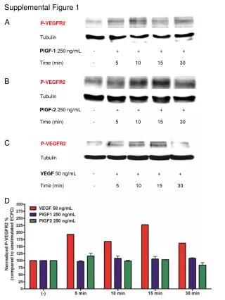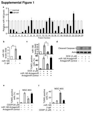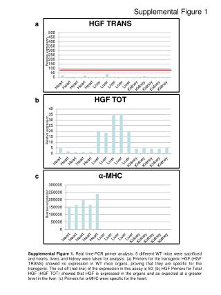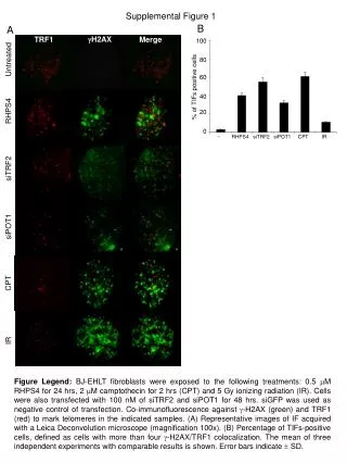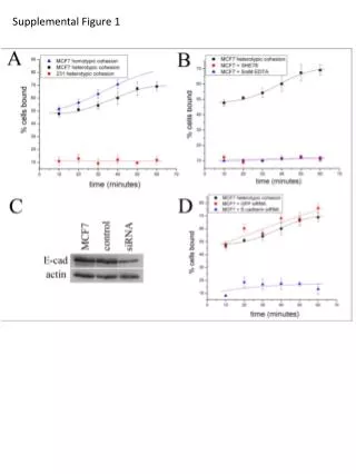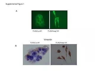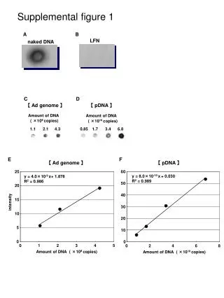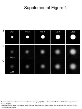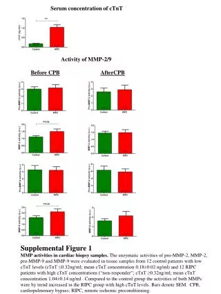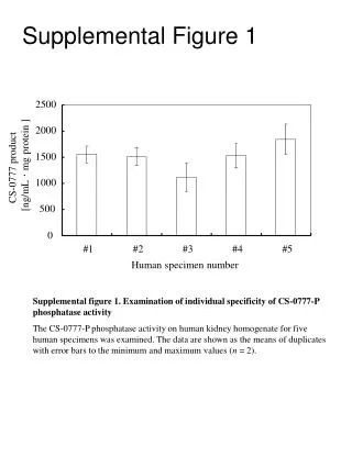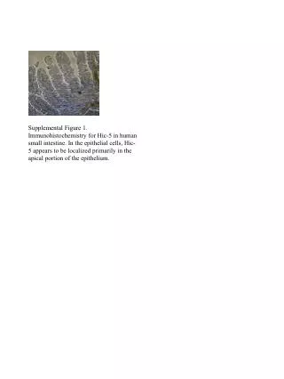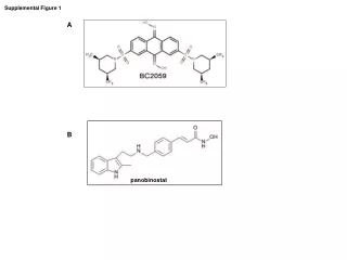
Supplemental Figure 1
20 likes | 204 Views
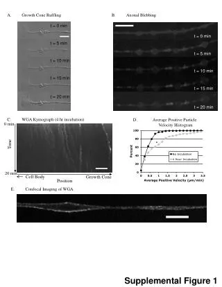
B. Axonal Blebbing. t = 0 min. t = 0 min. t = 5 min. t = 5 min. t = 10 min. t = 10 min. t = 15 min. t = 15 min. t = 20 min. t = 20 min. A. Growth Cone Ruffling. D. Average Positive Particle Velocity Histogram. C. WGA Kymograph (4 hr incubation). 0 min. Time. *. 20 min.

Supplemental Figure 1
E N D
Presentation Transcript
B. Axonal Blebbing t = 0 min t = 0 min t = 5 min t = 5 min t = 10 min t = 10 min t = 15 min t = 15 min t = 20 min t = 20 min A. Growth Cone Ruffling D. Average Positive Particle Velocity Histogram C. WGA Kymograph (4 hr incubation) 0 min Time * 20 min Cell Body Growth Cone Position E. Confocal Imaging of WGA WGA Supplemental Figure 1
A. Average Positive Particle Velocity Histogram B. Average Positive Particle Velocity Histogram (within 30 µm of the edge) D. Average Negative Particle Velocity Histogram (within 30 µm of the edge) C. Average Negative Particle Velocity Histogram Supplemental Figure 2