Download
1 / 27
1.58k likes | 6.69k Views
Clavicle Fractures. Arezu Jafarian Yazdi. What is Clavicle.

E N D
Clavicle Fractures Arezu Jafarian Yazdi
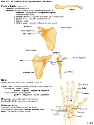
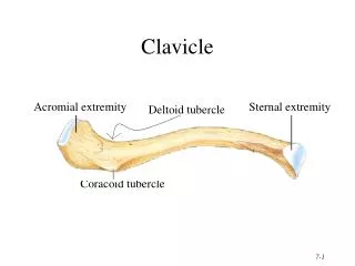
What is Clavicle Also known as the collarbone, a thin, slightly S-shaped bone attached by ligaments to the top of the sternum(breastbone). The clavicle is present in most vertebrates. In humans, the right and left clavicles and right and left scapulas (shoulder blades) make up the shoulder girdle, linking the arms to the axis of the body. The clavicle lies horizontally and articulates with the sternum and first costal cartilage medially and with the acromion of the scapula laterally. The clavicle acts as a strut holding the arm away from the trunk. It also serves to transmit forces from the upper limb to the axial skeleton, and provides attachment for muscles. The clavicle is subcutaneous throughout its length; its medial two-thirds are convex forward and its lateral third concave forward.
Epidemiology Clavicle fractures involve approximately 5% of all fractures seen in hospital emergency admissions. Clavicles are the most common broken bone in the human body. It is most often fractured in the middle third of its length which is its weakest point. The lateral fragment is depressed by the weight of the arm and is pulled medially and forward by the strong adductor muscles of the shoulder joint, especially the pectorals major. The part of the clavicle near the center of the body is tilted upwards by the sternocleidomastoid muscle. Children and infants are particularly prone to it. Newborns often present clavicle fractures following a difficult delivery.
Risk Factors/Prevention Those who have a low dietary intake of calcium and Vitamin D may have a higher risk of clavicular fractures. Increasing the integrity of the bone by a sufficient amount of dietary calcium and vitamin D will help to prevent fractures in the bone. Also, sedentary individuals may be at a higher risk due to weakness in muscle stabilizers of the clavicle. Also, participation in extreme sports such as mountain biking and snowboarding will increase the risk of a clavicular fracture as well.
Clavicle Fractures Clavicular fractures are classified mechanistically and anatomically into 3 types. Approximately 80% of clavicle fractures occur in the middle third (class A), 15% involve the distal or lateral third (class B), and less than 5% involve the proximal or medial third (class C). The anatomy of the clavicle with potential fracture sites marked is shown in the image below.
Fractures; • Proximal Clavicle Fractures [Type A] • Occurs in 5% of clavicle fractures making it a very uncommon injury to treat • The proximal clavicle fracture tends not to displace related to good ligamentous support. • Displaced proximal frvacturesrequire close examination for neurovascular compromise, and generally require reduction. • Nondisplaced proximal fractures are treated with sling for comfort, analgesia, and range of movement exercises when pain allows.
fractures • Midshaft Clavicle Fractures: [Type B] • Occurs in 80% of clavicle fractures, making it the most common type off fracture. • Diagnosis of midshaft clavicle fractures is generally straight forward based on history, examination and radiology. • Treatment of midshaft clavicle fractures involves restore normal anatomy, limit pain, and promote quick return to activity or play.
fractures • Distal Clavicle Fractures: [Type C] • Occurs in 15% of clavicle fractures • Distal clavicle fracture occurs distal to the coracoclavicular ligaments. • Divided into 3 types nondisplaced, displaced and articluar. • Displaced distal clavicle fractures tend to require internal fixation for support.
Signs and symptoms • Pain that increases with shoulder movement • Swelling • Tenderness • Bruising • A bulge on or near your shoulder • A grinding or crackling sound when you try to move your shoulder • Stiffness or inability to move your shoulder
Signs and symptoms • You may have pain, tenderness, swelling, bruising, or a bump in the injured area. • The bones may poke through the skin, not look normal, or look out of place. • The shoulder and arm may feel weak, numb, and tingly. • You may have trouble moving your shoulder and arm. • You may need to support the arm with your other hand to decrease the pain.
How is a clavicle fracture diagnosed • During the evaluation, your doctor will ask questions about the injury and how it occurred. After discussing the injury and your symptoms, your doctor will examine your shoulder. • There is usually an obvious deformity, or "bump," at the fracture site. Gentle pressure over the break will bring about pain. Although a fragment of bone rarely breaks through the skin, it may push the skin into a "tent" formation.
How is a clavicle fracture diagnosed • Computerized tomography scan: This is also called a CT or CAT scan. A special x-ray machine uses a computer to take pictures of your clavicle. You may be given a dye before the pictures are taken. The dye is usually given in your vein (IV). The dye may help your caregiver see the pictures better. People who are allergic to iodine or shellfish may be allergic to some dyes. Tell your caregiver if you are allergic to shellfish, or have other allergies or medical conditions.
How is a clavicle fracture diagnosed • Magnetic resonance imaging scan: This is also called an MRI. An MRI uses magnetic waves to take pictures of your clavicle, chest bone, and shoulder areas. During an MRI, pictures are taken of your bones, muscles, joints, or blood vessels. You will need to lie still during an MRI. Never enter the MRI room with an oxygen tank, watch, or any other metal objects. This may cause serious injury. • X-rays: You may need x-rays of your clavicle, chest bone, and shoulder to check for broken bones or other problems. X-rays of both your injured and uninjured clavicles may be taken.
Treatment Nonsurgical Treatment If the broken ends of the bones have not shifted out of place and line up correctly, you may not need surgery. Broken collarbones can heal without surgery. Arm Support A simple arm sling or figure-of-eight wrap is usually used for comfort immediately after the break. These are worn to support your arm and help keep it in position while it heals. Medication Pain medication, including acetaminophen, can help relieve pain as the fracture heals.
Nonsurgical Treatment • Physical Therapy • While you are wearing the sling, you will likely lose muscle strength in your shoulder. Once your bone begins to heal, the pain will decrease and your doctor may start gentle shoulder and elbow exercises. These exercises will help prevent stiffness and weakness. More strenuous exercises can gradually be started once the fracture is completely healed. • Doctor Follow-Up • You will need to see your doctor regularly until your fracture heals. He or she will examine you and take x-rays to make sure the bone is healing in good position. After the bone has healed, you will be able to gradually return to your normal activities. .
Nonsurgical Treatment Complications The fracture can move out of place before it heals. It is important to follow up with your doctor as scheduled to make sure the bone stays in position. If the fracture fragments do move out of place and the bones heal in that position, it is called a "malunion." Treatment for this is determined by how far out of place the bones are and how much this affects your arm movement. A large bump over the fracture site may develop as the fracture heals. This usually gets smaller over time, but a small bump may remain permanently
Surgical Treatment If your bones are out of place (displaced), your doctor may recommend surgery. Surgery can align the bones exactly and hold them in good position while they heal. This can improve shoulder strength when you have recovered. Plates and Screws During this operation, the bone fragments are first repositioned into their normal alignment, and then held in place with special screws and/or by attaching metal plates to the outer surface of the bone. After surgery, you may notice a small patch of numb skin below the incision. This numbness will become less noticeable with time. Because there is not a lot of fat over the collarbone, you may be able to feel the plate through your skin. Plates and screws are usually not removed after the bone has healed, unless they are causing discomfort. Problems with the hardware are not common, but sometimes, seatbelts and backpacks can irritate the collarbone area. If this happens, the hardware can be removed after the fracture has healed.
Surgical Treatment (A) The clavicle is broken in more than one place and the fragments are severely out of alignment. (B)The fractured pieces are held in place by a combination of plates and screws.
SurgicalTreatment Pins Pins are also used to hold the fracture in good position after the bone ends have been put back in place. The incisions for pin placement are usually smaller than those used for plates. Pins often irritate the skin where they have been inserted and are usually removed once the fracture has healed. Rehabilitation Specific exercises will help restore movement and strengthen your shoulder. Your doctor may provide you with a home therapy plan or suggest that you work with a physical therapist. Therapy programs typically start with gentle motion exercises. Your doctor will gradually add strengthening exercises to your program as your fracture heals. Although it is a slow process, following your physical therapy plan is an important factor in returning to all the activities you enjoy.
Clavicle Fractures ORIF Displaced
Clavicle Fractures Distal clavicle plate
Clavicle Fractures IM Rod
SurgicalTreatment • Surgical Complications • People who use any kind of tobacco product, have diabetes, or are elderly are at a higher risk for complications during and after surgery. They are also more likely to have problems with wound and bone healing. Be sure to talk with your doctor about the risks and benefits of surgery for your clavicle fracture. • There are risks associated with any surgery, including: • Infection • Bleeding • Pain • Blood clots in your leg • Damage to blood vessels or nerves • Nausea • The risks specific to surgery for collarbone fractures include: • Difficulty with bone healing • Lung injury • Hardware irritation