Download
1 / 62
620 likes | 628 Views
Biology 212 Anatomy & Physiology. Respiratory System. Functions of Respiratory System Ventilation : Movement of air to and from sites of gas exchange Gas Exchange : Movement of specific gases such as oxygen & carbon dioxide between air and

E N D
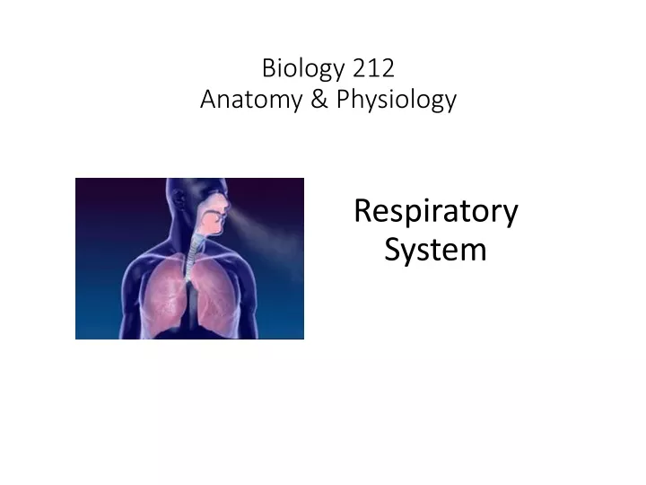
Biology 212Anatomy & Physiology Respiratory System
Functions of Respiratory System Ventilation: Movement of air to and from sites of gas exchange Gas Exchange: Movement of specific gases such as oxygen & carbon dioxide between air and blood (in lung) or between blood and extracellular fluids (in other tissues) Gas Transport: Movement of oxygen (in blood) away from lung and of carbon dioxide (also in blood) back toward lung. Obviously: Closely linked with circulatory system
Organs of Respiratory System Nasal Cavity Pharynx Larynx Trachea Bronchi Lungs AirBlood Smaller bronchi Branches of pulmonary arteries Bronchioles Arterioles Alveolar ducts Capillaries Alveoli Venules Branches of pulmonary veins
Nasal Cavity: Removes dust & other debris Warms inhaled air Humidifies inhaled air Concha or turbinates project from lateral wall, create turbulance
Right & left sides of nasal cavity separated by nasal septum; Nasal cavity separated from oral cavity by palate
Inhaled air passes from nasal cavity into pharynx.
Inhaled air passes from nasal cavity into pharynx. Three regions: Nasopharynx
Inhaled air passes from nasal cavity into pharynx. Three regions: Nasopharynx Oropharynx
Inhaled air passes from nasal cavity into pharynx. Three regions: Nasopharynx Oropharynx Laryngopharynx Inhaled air passes from laryngopharynx into larynx
Inhaled air passes from nasal cavity into pharynx. Three regions: Nasopharynx – Pseudostratified columnar epithelium Oropharynx Mixture of pseudostratified columnar Laryngopharynx and stratified squamous epithelia
Functions of Larynx: - Keeps airway open even with negative pressure - Keeps food / liquids from entering trachea - Vocalization
Structure of Larynx: Nine cartilages connected by muscles & ligaments
Structure of Larynx: Nine cartilages: 3 large unpaired: Thyroid Cricoid Epiglottis 6 smaller paired: Arytenoid Corniculate Cuneiform Anterior View Posterior View
Anterior Posterior Midsagittal (Section) Superior
Vocal cords (also called vocal folds or vocal ligaments) are strands of dense regular connective tissue running anteriorly from arytenoid cartilages to thyroid cartilage. If they are adducted (close together) air moving between the cords causes them to vibrate.
Vocal cords (also called vocal folds or vocal ligaments) are strands of dense regular connective tissue running anteriorly from arytenoid cartilages to thyroid cartilage. Air moving between them cause them to vibrate. Intrinsic muscles of larynx (no, you don’t need to know their names) move the vocal cords by moving the arytenoid cartilage Opening between vocal cords = glottis
Trachea: Begins at bottom of larynx (cricoid cartilage) Ends by dividing into two primary bronchi. Cricoid Cartilage Primary Bronchus
Trachea: Held open by cartilages which form incomplete rings around it. Lumen lined by pseudostratified columnar epithelium. Many mucous glands in wall; cilia on surface
Trachea: Cross section of neck at level of vertebra cervical 6
Lungs: Occupy most of thoracic cavity
Lungs: Primary bronchus, pulmonary artery, and pulmonary veins enter/leave lung together at hilum (or hilus or root) of each lung
Lungs: Surrounded by double-layered pleura with pleural cavity between parietal and visceral layers
Primary bronchus Pulmonary artery Pulmonary vein
Lungs: Each lung divided into lobes by deep grooves or fissures Right lung has 3 lobes: Superior lobe Middle lobe Inferior lobe
Lungs: Each lung divided into lobes by deep grooves or fissures Right lung has 3 lobes: Superior lobe Middle lobe Inferior lobe Separated by 2 fissures: Horizontal fissure Oblique fissure
Lungs: Each lung divided into lobes by deep grooves or fissures Left lung has 2 lobes: Superior lobe Inferior lobe
Lungs: Each lung divided into lobes by deep grooves or fissures Left lung has 2 lobes: Superior Inferior Separated by 1 fissure: Oblique fissure
Lungs: Each lung divided into lobes by deep grooves or fissures Each lobe has is own secondary bronchus, its own branch of the pulmonary artery, and its own branch of the pulmonary vein
Lungs: Each lobe consists of bronchopulmonary segments Each segment has is own tertiary bronchus, its own branch of the pulmonary artery, and its own branch of the pulmonary vein
Trachea Primary bronchi Secondary bronchi Tertiary bronchi (smaller branches) (bronchioles) Terminal bronchioles Respiratory bronchioles Alveolar ducts Alveoli
Trachea Primary bronchi Secondary bronchi Tertiary bronchi (smaller branches) (bronchioles) Terminal bronchioles Respiratory bronchioles Alveolar ducts Alveoli Conducting Zone Respiratory Zone
Alveoli: Microscopic air sacs Diameter 200-400 um Wall = simple squamous epithelium called Type I Alveolar cells Other cells: Type II alveolar cells (secrete surfactant) Dust cells (macrophages)
As the bronchi branch and divide, so do the pulmonary arteries and pulmonary veins which accompany them. At the end, each alveolus is surrounded by many capillaries for the exchange of gasses between air (in the alveolus) and blood (in the capillaries).
This air (in the alveolus) and blood (in the capillaries) are separated by a very thin wall, called the respiratory membrane, through which gasses can easily diffuse.
Let’s return to ventilation: Humans inhale by creating a negative pressure (or suction) in the lungs. Movement of the diaphragm and intercostal muscles cause the thoracic cavity to get larger. The lung expands to fill this larger space, which lowers the pressure of the air within the alveoli. Since the pressure of the air in alveoli is now lower than the pressure of atmospheric air, it gets pushed from the mouth and nose through the pharynx, larynx, trachea, bronchi, bronchioles, and alveolar ducts into the alveoli.
Air pressure is measured according to how hard it can push on other substances. The most common way is to measure how far it can push mercury (a liquid metal with the chemical symbol Hg) up a tube. If it can push mercury 750 millimeters (ml) up a tube, we say the air pressure is “750 millimeters of mercury”, abbreviated “750 mm Hg”.
Air pressure is measured according to how hard it can push on other substances. The most common way is to measure how far it can push mercury (a liquid metal with the chemical symbol Hg) up a tube. If it can push mercury 750 millimeters (ml) up a tube, we say the air pressure is “750 millimeters of mercury”, abbreviated “750 mm Hg”. Here’s why this is important: Air will always move from the place with higher pressure to the place with lower pressure
Terminology you need to know: Atmospheric pressure Intrapulmonary pressure (pressure of air in alveoli) Intrapleural pressure (pressure of air in pleural cavity) Difference between intrapleural pressure and intrapulmonary pressure is called transpulmonary pressure
Proper ventilation requires that the lungs also expand each time the thoracic cavity expands. This can not happen if air enters the pleural cavity (pneumothorax) or if blood enters the pleural cavity (hemothorax). These cause the lung (covered by visceral pleura) to collapse and pull away from the chest wall (lined by parietal pleura). Called atalectasis.
Respiratory Volumes: TIDAL VOLUME: The volume of air which moves in and out of the lungs with a normal breath (normal = 400-500 ml)
Respiratory Volumes: EXPIRATORY RESERVE VOLUME: The volume of air, beyond tidal volume, which can be forcibly expired (normal = + 1,200 ml)
Respiratory Volumes: INSPIRATORY RESERVE VOLUME: The volume of air, beyond tidal volume, which can be forcibly inhaled (normal = + 3,000 ml)
Respiratory Volumes: RESIDUAL VOLUME: The volume of air which remains in the lungs after forcible expiration (normal = + 1,200 ml)
Respiratory Volumes: VITAL CAPACITY: = Sum of Tidal volume + Inspiratory reserve volume + Expiratory reserve volume
Respiratory Volumes: TOTAL LUNG CAPACITY: = Tidal volume + Inspiratory reserve volume + Expiratory reserve volume + Residual volume
Note that all of the air which enters your nose does not reach your alveoli. Approximately 150 ml of air remains in the nasal cavity, pharynx, larynx, trachea, bronchi, and bronchioles. This = Anatomical dead space.
Gas Exchange: Movement of specific gases: • From a mixture of gases into • a liquid (e.g. oxygen moves • from air in the alveoli into • blood in the capillaries) • From a liquid into a mixture • of gases (e.g. carbon dioxide • moves from blood in the • capillaries into air in the alveoli) • From one liquid into another liquid (e.g. oxygen leaves the blood and diffuses into extracellular fluid, while carbon dioxide moves from the extracellular fluids into the blood.