Download
1 / 37
500 likes | 1.12k Views
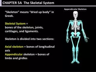
CHAPTER 5A The Skeletal System. Appendicular Skeleton. Axial Skeleton. “Skeleton” means “dried up body” in Greek. Skeletal System = bones of the skeleton, joints, cartilages, and ligaments. Skeleton is divided into two sections: Axial skeleton = bones of longitudinal axis

E N D
CHAPTER 5A The Skeletal System Appendicular Skeleton Axial Skeleton “Skeleton” means “dried up body” in Greek. Skeletal System = bones of the skeleton, joints, cartilages, and ligaments. Skeleton is divided into two sections: Axial skeleton = bones of longitudinal axis Appendicular skeleton = bones of limbs and girdles
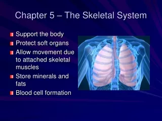
CHAPTER 5A The Skeletal System • Functions of Bones • Support (internal framework) • Protection (of organs) • Movement (with skeletal muscles) • Storage (fat, minerals – especially calcium and phosphorus) • Calcium comes and goes from bones continually as it is needed in the blood. • Blood Cell Formation (hematopoiesis) occurs in bone marrow
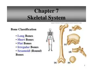
CHAPTER 5A The Skeletal System • Classification of Bones • Compact Bone = dense, smooth, homogeneous • Spongy Bone = made of small needlelike pieces of bone and open spaces
CHAPTER 5A The Skeletal System • Classification of Bones • Long Bones • Longer than wideHead at each endMostly compact bone • Limb bones
CHAPTER 5A The Skeletal System • Classification of Bones • Short Bones • Cube-shapedSpongy bone • Ex. Wrist and ankle
CHAPTER 5A The Skeletal System • Classification of Bones • Flat Bones • Thin, flattened, usually curved. • Two thin layers of compact bone sandwiching spongy bone. • Ex. Skull, ribs, and sternum.
CHAPTER 5A The Skeletal System • Classification of Bones • Irregular Bones • Don’t fit into other categories. • Ex. Vertebrae, hip bones.
CHAPTER 5A The Skeletal System Structure of a Long Bone Gross Anatomy (See TXT pg 114 Fig 5.2) Diaphysis = shaft Periosteum = covers shaftSharpy’s Fibers = hold periosteum to bone Epiphyses = ends of boneArticular cartilage = covers epiphyses Epiphyseal line = only in adults Epiphyseal plate = growth portion in youth
CHAPTER 5A The Skeletal System Structure of a Long Bone Gross Anatomy (See TXT pg 114 Fig 5.2) Red Marrow Infants: forms blood cells Adults: only in spongy bone of flat bones. Yellow Marrow Adults only.
CHAPTER 5A The Skeletal System Structure of a Long Bone Gross Anatomy (See TXT pg 115 Table 5.1) Bone Markings Categories: Projections (Processes) Grows out from bone surface. Depressions (Cavities) Indentations into the bone.
CHAPTER 5A The Skeletal System Microscopic Anatomy (See TXT pg 116 Fig 5.3) Canals connect all tissues Circles of lacunae Lamellae surround it Central canal + matrix rings Communication pathway Cavities in matrix Mature bone cells
CHAPTER 5A The Skeletal System Bone Growth (See TXT pg 117 Fig 5.4) For this slide, you will receive a handout that accompanies the skeletal system interactive video. Vocabulary will be included in the handout. Skeletal System Interactivehttp://player.discoveryeducation.com/index.cfm?guidAssetId=B7D2D569-417F-454B-8490-4681832950A5&blnFromSearch=1&productcode=US Bone Growth http://highered.mcgraw-hill.com/sites/0072507470/student_view0/chapter6/animation__bone_growth_in_width.html
CHAPTER 5A The Skeletal System Fractures (See TXT pg 119 Table 5.2) Types of Bone Fractureshttp://video.about.com/orthopedics/Fractures-2.htm
CHAPTER 5A The Skeletal System Fractures More Fractures online… Click-on Fracture Animations http://www.argosymedical.com/Skeletal/index.html
CHAPTER 5A The Skeletal System Fractures Reduction = repair of fractures. Emergency procedures for broken bones http://www.argosymedical.com/Skeletal/samples/animations/Broken%20Bone/index.html
CHAPTER 5A The Skeletal System Fractures
CHAPTER 5A The Skeletal System / Bones (See TXT pg 120 Fig 5.5) • Stages of Healing: • Hematoma • Soft Callus • Bony Callus • Remodeling
CHAPTER 5A The Skeletal System / Bones (See TXT pg 121 Fig 5.6) Axial Skeleton: Skull Vertebral column Bony thorax
CHAPTER 5A The Skeletal System / Bones (See TXT pg 122 Fig 5.7) The SKULL Laterial View of the Skull
CHAPTER 5A The Skeletal System / Bones Ancient man following skull surgery. Other Lateral Views of Skull This weird looking skull is the famous Starchild Skull that was found in 1930. The Starchild Skull was discovered in a mine tunnel about 100 miles southwest of Chihuahua, Mexico along with several other normal skeletons. 100,000-year-old skull was found in a cave in Qafzeh, Israel,
CHAPTER 5A The Skeletal System / Bones Anterior View of the Skull
CHAPTER 5A The Skeletal System / Bones More Anterior Views of Skulls 8-13 yr old boy
CHAPTER 5A The Skeletal System / Bones Sinus = air filled cavity Process = projection Foramen = hole or opening Suture = immovable fibrous joints between bones Condyle = rounded projection at the end of a bone that articulates with another bone Paranasal Sinuses
CHAPTER 5A The Skeletal System / Bones Hyoid Bone Not part of skull. Only bone that does not interact directly with another bone. Suspended and anchored by ligaments. Moveable base for tongue. Attachment point for swallowing. Aids in speech.
CHAPTER 5A The Skeletal System / Bones Fetal Skull Face is small compared to cranium. Skull overall is large for the body. Cranial bones connected by fontanels which harden post partem. http://ect.downstate.edu/courseware/haonline/quiz/practice/u5/quiztop5.htm
CHAPTER 5A The Skeletal System / Bones Vertebral Column Also called spine. Before birth = 33 vertebrae After birth = 24 vertebrae
CHAPTER 5A The Skeletal System / Bones Vertebral Column Separated into 5 major sections: Cervical (neck) Thoracic (mid back) Lumbar (lower back) Sacrum (top of tail) Coccyx (tail bone) Scoliosis Animation http://www.spine-health.com/video/scoliosis-video-what-scoliosis
CHAPTER 5A The Skeletal System / Bones Vertebral Column Intervertebral discs = separate vertebrae 90% water Dries as ages. Herniated discs results from drying and weakening of ligaments holding discs in place. Herniated disc animation http://www.spine-health.com/video/lumbar-herniated-disc-video
CHAPTER 5A The Skeletal System / Bones Vertebral Column S-shaped structure Provides flexibility and prevents shock to the brain Primary curvatures formed at birth Thoracic and Sacral Secondary curvatures cervical – when baby begins raising head lumbar – when baby begin walking
CHAPTER 5A The Skeletal System / Bones Vertebral Foramen Body Weight bearing Pedicle Superior Articular Process Transverse Process Vertebral Arch Lamina Thoracic Vertebra Spinous Process
CHAPTER 5A The Skeletal System / Bones 1st Vertebra: Atlas Has no body Holds occipital condyles of skull Allows nodding = “yes” 2nd Vertebra: Axis Acts as pivot for rotation of skull Allows side-to-side motion = “no” Dens: Pivot point Cervical Vertebra
CHAPTER 5A The Skeletal System / Bones Thoracic Vertebra Supports ribs.
CHAPTER 5A The Skeletal System / Bones Lumbar Vertebra Block-like body. Most of stress on spine occurs in lumbar region.
CHAPTER 5A The Skeletal System / Bones Sacrum Formed by fusion of 5 vertebrae. Alae = articulates with hip bones. Formed by fusion of 3 vertebrae. “Tailbone” Coccyx
CHAPTER 5A The Skeletal System / Bones Bony Thorax Ribs: True Ribs: 1st 7 ribs Attach to sternum. False Ribs: Last 5 ribs Attach indirectly to sternum or not attached at all. Last 2 false ribs are “floating ribs” – unattached.
CHAPTER 5A The Skeletal System / Bones Bony Thorax Sternum: Manubrium ProcessBody Xiphoid process easily broken during CPR
CHAPTER 5A The Skeletal System / Bones TEST TIME! CH 5 AXIAL SKELETON