Download
1 / 47
610 likes | 1.17k Views
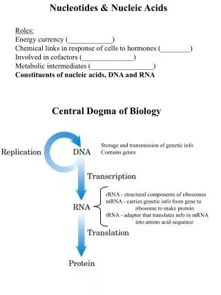
Introduction to Nucleotides and Nucleic Acids. Chapter 8 (Page 273-283). Fundamental Biology of Life. Nucleus: Site of transcription. DNA RNA Protein. Ribosomes on endoplasmic reticulum: Site of translation. Transcription. Translation.

E N D
Introduction to Nucleotides and Nucleic Acids Chapter 8 (Page 273-283)
Fundamental Biology of Life Nucleus: Site of transcription DNA RNA Protein Ribosomes on endoplasmic reticulum: Site of translation Transcription Translation Replication
1. Nucleotides- General Function Participate in: • Energy exchange processes in metabolism (ATP) • Metabolic intermediates • Cellular response to extracellular stimuli and intracellular signal transduction (cAMP) • Structural components of enzyme and enzyme cofactors (NAD+) • The constituents of nucleic acids, the molecular repositories of genetic information I. Deoxyribonucleic acid (DNA) II. Ribonucleic acid (RNA)
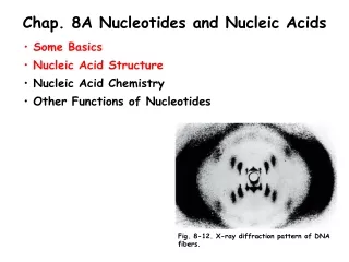
2. Nucleotide Composition Have three characteristic components • Nucleobase (Nitrogen-containing base) • Pentose • Phosphate *
2I. Nucleobase • Derivatives of pyrimidine or purine B. Nitrogen-containing heteroaromatic molecules C. Planar or almost planar structures D. Absorb UV light around 250–270 nm * *
2IA. Pyrimidine Bases 4 5 3 2 6 1 * * *
2IA. Pyrimidine Bases A. Cytosine is found in both DNA and RNA Thymine is found only in DNA Uracil is found only in RNA B. All are good H-bond donors and acceptors C. Cytosine pKa at N3 is 4.5 Thymine pKa at N3 is 9.5 D. They are neutral molecules at pH 7
2IB. Purine Bases 6 7 1 5 8 2 4 9 3 * *
2IB. Purine Bases • Adenine and guanine are found in both RNA and DNA B. Also good H-bond donors and acceptors C. Adenine pKa at N1 is 3.8 Guanine pKa at N7 is 2.4 D. Neutral molecules at pH 7
2IC. Tautomerism of Nucleobases The nucleobases can exist in different structural isomeric forms called prototropictautomers, which differ in the location of protons. The nucleobases undergo lactam-lactimtautomerism • The lactam form dominates at neutral pH
2ID. UV Absorption of Nucleobases The absorption of UV light at 250-270 nm by nucleobases is due to * electronic transitions. • These absorbances can be used to quantify nucleic acids.
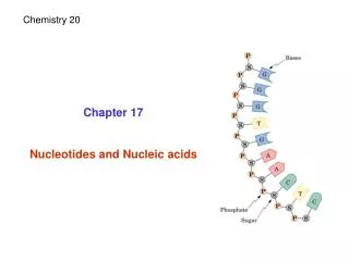
2II. Pentoses • The carbon numbers are given a prime (‘) designation to distinguish them from the numbered atoms of the nitrogenous bases. • Nucleic acids can have two kinds of pentoses. H Present in RNA Present in DNA β-2’-deoxy-D- ribofuranose β-D-ribofuranose D-ribose
2II. Pentoses C. The pentose ring is not planar but occurs in one of four “puckered” conformations. • Four of the five atoms are in a single plane. • The fifth atom is on either the same (endo) or the opposite (exo) side of the plane relative to the C-5’ atom.
2IIA. β-N-Glycosidic Bond In nucleotides the pentose ring is attached to the nucleobase via an N-glycosidic bond: • The bond is formed to the anomeric carbon of the sugar in β configuration • And to • Position N1 in pyrimidines • Position N9 in purines • This bond is quite stable toward hydrolysis, especially in pyrimidines • Bond cleavage is catalyzed by acid
2III. Phosphates • The phosphate group is typically esterified to the 5’ carbon of pentose. • The phosphate group can be in other positions. 2’-monophosphate 2’,3’-cyclic monophosphate 3’-monophosphate
3. Nucleoside Composition Have two of the three characteristic components of nucleotides • Nucleobase (Nitrogen-containing base) • Pentose
4. Nomenclature- Deoxyribonucleotides You need to know structures, names, and symbols.
4. Nomenclature- Ribonucleotides You need to know structures, names, and symbols.
5. Nucleic Acids can contain Unusual Nucleobases Modification of nucleobases is done after RNA/DNA synthesis: • 5-Methylcytosine is common in eukaryotes, also found in bacteria • N6-Methyladenosine is common in bacteria but not found in eukaryotes Modifications serve an epigenetic marker role: • Way to mark own DNA so that cells can degrade foreign DNA (prokaryotes); a defense mechanism • Way to mark which genes should be active (eukaryotes)
1. Nucleic Acids- General Function Nucleic acids are biologically occurring polynucleotides in which the nucleotide residues are linked in a specific sequence by phosphodiester bonds. They participate in: • Storage of genetic information (Deoxyribonucleic acid; DNA) • Transmission of genetic information (messenger ribonucleic acid; mRNA) • Processing of genetic information (ribozymes) • Protein synthesis (transfer RNA (tRNA) and ribosomal RNA (rRNA))
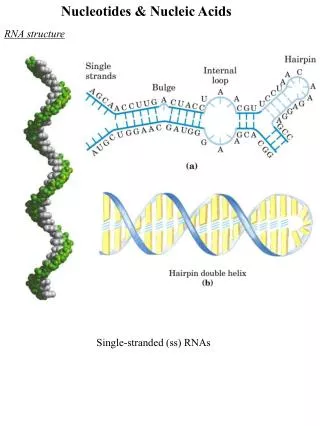
2. Levels of Nucleic Acid Structure • Primary Level • The nucleotide sequence of a nucleotide strand and its covalent structure • Secondary Level • Any regular, stable structure taken up by some or all of the nucleotides in a nucleic acid C. Tertiary Level • The complex folding of large chromosomes within eukaryotic chromatin (Chapter 24)
Primary Level of Nucleic Acid Structure
1. General Structure of Nucleotide Strands in Nucleic Acid The successive nucleotides of both DNA and RNA are covalently linked through phosphate-group “bridges,” in which the 5’-phosphate group of one nucleotide unit is joined to the 3’-hydroxyl group of the next nucleotide. • The covalent backbones of nucleic acids consist of alternating phosphate and pentose residues • The backbone is hydrophilic • The hydroxyl groups of the sugar residues form hydrogen bonds with water • The phosphate groups are completely ionized and negatively charged at pH 7
1. General Structure of Nucleotide Strands in Nucleic Acid • B. The nucleobases may be regarded as side groups • C. All phosphodiester linkages have the same orientation along the chain with distinct 5’ and 3’ ends. • D. The 5’ end lacks a nucleotide at the 5’ position and the 3’ end lacks a nucleotide at the 3’ position. • Other groups may be present on one or both ends. • E. The nucleotide sequences are arranged as linear polymers • No branching or cross-links
1. General Structure of Nucleotide Strands in Nucleic Acid • F. The DNA backbone is fairly stable • Hydrolysis accelerated by enzymes (DNAse) • DNA’s half-life is 521 years • DNA strands of a reasonable length could last 1 million years if preserved properly
1. General Structure of Nucleotide Strands in Nucleic Acid • G. The RNA backbone is unstable • In water, RNA lasts for a few years • In cells, mRNA is degraded in few hours
2. Hydrolysis of RNA • RNA is unstable under alkaline conditions • In the body hydrolysis is catalyzed by enzymes called RNases • Rnase P is a ribozyme (enzyme made of RNA) that processes tRNAprecurors • Dicer is an enzyme that cleaves double-stranded RNA into oligonucleotides (50 or fewer nucleotides) • - Helpful in defense against viral genomes
3. Representation of Nucleotide Sequences By convention , the sequence of a single strand of nucleic acid is always written with the 5’-end at the left and the 3’-end at the right. 5’ 3’ For a nucleic acid that consists of the AGCTA sequence, you can write the following : P = phosphate group; OH = 3’ end • pA-G-C-T-AOH • pApGpCpTpA • pAGCTA • 5’-AGCTA-3’
Secondary Level of Nucleic Acid Structure (DNA)
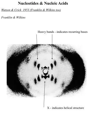
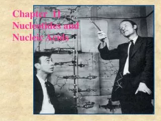
1. Discovery of DNA Structure • “This structure has novel features which are of considerable biological interest” • ―Watson and Crick, Nature, 1953 • One of the most important discoveries in biology • The pathway to discovery illustrates important factors about science • Missteps in modeling • Value of knowledge • Value of collaboration • Cost of sharing data too early
2. Covalent Structure of DNA (1868-1935) • Friedrich Miescher (1868) isolates “nuclein” from cell nuclei • Hydrolysis of nuclein - phosphate - pentose - and a nucleobase • Chemical analysis - phosphodiesterlinkages - pentose is ribofuranoside Structure of DNA: 1929 (Levene & London) Structure of DNA: 1935 (Levene & Tipson)
3. DNA Molecules have Distinctive Base Compositions • Erwin Chargaff and colleagues (late 1940s) found that the four nucleotide bases of DNA occur in different ratios in the DNAs of different organisms and that the amounts of certain bases are closely related. • The base composition of DNA generally varies from one species to another. • DNA isolated from different tissues of the same species have the same base composition. • In all cellular DNA • # of A = # of T • # of G = # of C • # of purines (A + G) = # of pyrimdines (T + C)
5. The Nobel Prize for Solving the Structure of DNA goes to…
6. Watson-Crick Model of DNA The DNA structure consists of: Right-handed double helix Hydrophilic backbone of alternating deoxyribose and phosphate groups faces outwards and interacts with H2O The furanose ring of each deoxyribose in the C-2’ endo conformation The purine and pyrimidine bases, which are hydrophobic and relatively insoluble in water, stacked inside the double helix stabilized by hydrophobic interactions and perpendicular to the long axis
6. Watson-Crick Model of DNA • E. Base pairing occurs in which a nucleobase in one strand is paired in the same plane with a base on the other strand due to hydrogen bond interactions. • Purine pairs with a pyrimidine • A pairs with T • C pairs with G • The base pairing of the two strands creates a major groove and a minor groove on the surface of the duplex
6. Watson-Crick Model of DNA • F. The DNA strands interact in an antiparallel orientation with the sequences being complementary to one another • 5’-ATGCTA-3’ • 3’-TACGAT-5’ • G. The stacked bases are 3.4 Å apart • H. Each turn of the helix (measured from one minor groove to the next minor groove) • Includes 10.5 base pairs stacked • Covers 36 Å