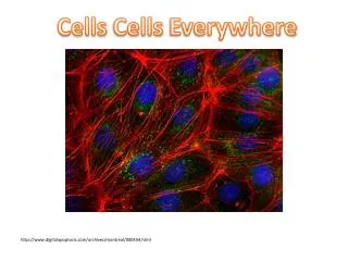
Cells
Methods in cell biology and functional genomics overview. Individualized. High throughput. FACS. Cells. DNA. FACS. Genomics. Markers. RNA. Tissues. Transcriptomics. In situ hyb. Protein. Organs. Immuno techniques. Proteomics. Metabolite. Organisms. Microscopy. Metabolomics.


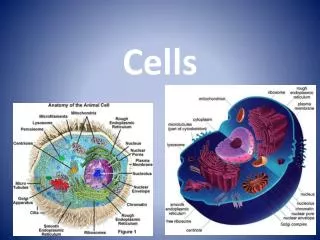
Cells
E N D
Presentation Transcript
Methods in cell biology and functional genomics overview Individualized High throughput FACS Cells DNA FACS Genomics Markers RNA Tissues Transcriptomics In situ hyb Protein Organs Immuno techniques Proteomics Metabolite Organisms Microscopy Metabolomics Structure Wild type Mutant
Antibody Purification • To reduce background, antibodies need to be purified before use. • Common procedures: • Ammonium sulphate precipitation. • Affinity chromatography a. Ammonium sulphate (40% v/v) precipitation is used to purify immunoglobulins from a large volumes of liquids. Precipitated proteins are collected by centrifugation, dissolved in PBS, dialyzed against PBS, and filtered before use. Disadvantage: all proteins from the serum are precipitated; may or may not remove the proteins causing background problems.
Affinity Chromatography Makes use of specific binding interactions between molecules 1- Incubate crude sample with the immobilized ligand 3- Elute 2- Wash away non-bound sample components from the solid support
Affinity Chromatography • Protein A (Staphylococcus aureus) and Protein G (Streptococcus sp.) bind to the Fc portion of immunoglobulins from many species. • Protein A binds to Fc- γ and Fv-VHIII . • Protein G binds to Fc- γ and c- γ1 chains. • Many monoclonal antibodies, and expressed fragments of antibodies containing only the antigen-binding site, do not bind to protein A and protein G. • Solution: A molecule with broader Ig-binding activity, including affinity for Fab fragments of the different classes of Ig can be used. • Protein L, binds Ig molecules regardless of heavy chain class, through interaction with Ig light chains. Ref: Hilson et al. (1993): Journal of Immunological Methods, 164:33-40.
Affinity Chromatography • Commonly used elution strategies for affinity chromatography: • pH • Ionic strength • Denaturation • Competition using ligand or analog For antibody purification, a combination of high salt and low pH is typically used to elute the antibodies.
Applications using Monoclonal and Polyclonal Antibodies • Western Blotting • Immunoprecipitation • Immunocytochemistry • Immuno electron microscopy • Immunofluorescence • Enzyme-Linked Immunosorbent Assay (ELISA) • Chromatin IP (ChIP) • Flow Cytometry (FC)
Western Blotting • Technique to specifically identify your favorite antigen extracted from biological samples. What it tells you: 1. The size of a protein. 2. The amount of protein present (semi-quantitative). 3. In what tissue a protein is expressed. What it doesn’t tell you (without further evidence): 1. Whether the protein normally exists as part of a larger complex. 2. Subcellular localization of the protein
Western Blotting • Sample separated using gel electrophoresis
Western Blotting Incubate membrane with primary antibody. Wash. Incubate membrane with secondary antibody that is radioactive or conjugated to an enzyme that generates light, either of which can be seen on a film, or simply by a color change (depending on what is conjugated to the secondary antibody). Same principle is used for the immunofluorescence techniques to be described a few slides later.
Monoclonal Antibody Specificity • The ACT1 peptide successfully competes with the MAb45a monoclonal antibody, indicating it is the epitope recognized by this anti-actin1 antibody. • The ACT11 peptide does not compete (this differs from ACT1 by only one amino acid (red arrow). Ref: Kandasamy et al. (1999). Plant Journal 164:33-40.
Immunocytochemistry • Technique used to label your favorite antigen within a cell and visualize it using fluorescence microscopy. Red = antibody to actin Blue = antibody to a nuclear protein Green = antibody to a Golgi protein Pink = antibody to a mitochondrial protein NIH 3T3 cells
Immunocytochemistry What it tells you: Shows subcellular localization of proteins. Which cells express that protein. May suggest an interaction. What it doesn’t tell you: Very difficult to quantify.
Immunocytochemistry Fix tissues, impregnate with resin/paraffin, section tissues. Remove resin/paraffin and add antibody. Direct labeling: antibody conjugated with detection tags. Indirect labeling: Add secondary antibody which is coupled to a detection tag. Disadvantage: Fixation artifacts, cells are dead at the time of detection.
Immunocytochemistry - Use of antibodies raised against a small molecule Tissues (wild type or mutant) sections probed with anti-GABA monoclonal antibodies B,F: Detection using secondary antibody conjugated to HRP detected with silver. C,D,G,H: Detection with secondary antibody conjugated to TRITC. Palanivelu et al., (2003). Cell 114:47-59.
Immunoprecipitation • Technique used to purify antigen from out of a complex mixture. Analyze complex
Protein A Protein B Antibody to protein B bound to beads G Co-Immunoprecipitation (Co-IP) • Technique used to investigate protein-protein interactions and isolate protein complexes. Immunoprecipitate with an antibody to A 2. Check pellet fraction to see if it had brought down protein B with an antibody to protein (or using gel electrophoresis, or mass spectrometry). 3. If no antibody to protein B is available, use radiolabeled or tagged version of protein B. 4. Does not prove the two proteins interact directly; they may be part of a larger complex.
Enzyme-Linked Immunosorbent Assay (ELISA) • Technique used to detect presence of proteins within a solution and quantify their amounts. • Used to detect: • Presence and quantity of an antigen or antibody. • Efficiency of antigen extraction from cells. • Examples • Tuberculosis test. • HIV test. • Pregnancy test.
Resolution is limited by the wavelength of radiation Nondestructive Live imaging Challenge: sample preparation Computerized 3-D reconstruction
Probe these sections with antibodies; Electron-dense label (ferritin • or colloidal gold) is conjugated to the Fc portion.
electron-dense labels absorbs electrons. • An immunoelectronmicrograph of the surface of a B-cell lymphoma was stained with two antibodies (Ab against class II MHC labeled sith 30nm gold particles, & another Ab against class I MHC w/ 15nm gold particles. • (The density of class I exceeds that of class II)
Chromatin Immunoprecipitation (ChIP) Formaldehyde crosslinks DNA to protein, capturing in vivo contacts. Shear chromatin , and perform immunoprecipitationwith specific antibody. Reverse crosslinks. Analyze recovered DNA.
Chromatin Immunoprecipitation (ChIP) • Types of antibody used in ChIP: • Recognizing histones, or various histone modifications (acetylation, methylation, phosphorylation). • Recognizing components of the transcription machinery. • Recognizing specific TFs. • Recognizing methylated DNA (cf. methyl cytosine).
ChIP Readouts • Microarrays (ChIP-chip). Primarily using promoter arrays or tiling arrays. • Sequencing (ChIP-seq). Originally employed SAGE methods. Best current platform is Solexa Next Generation sequencing.
ChIP Readouts • Microarrays (ChIP-chip). • Sequencing (ChIP-seq).
ChIP Readouts • Zhang et al. (2006). Genome-wide high-resolution mapping and functional analysis of DNA methylation in Arabidopsis. Cell 126:1189-1201. • Used a monoclonal antibody directed against methyl cytosine (mCIP), as well as the methyl cytosine binding domain (MBD) from the human protein MeCP2, as the affinity tags.
Euchromatic region Centromeric region FWA Note effects of ddc mutant (eliminates non-CG methylation) and met1 (eliminates all CG methylation and substantial amount of non-CG methylation)
ChIP-seq Readouts • Johnson DS, Mortazavi A, Myers RM, Wold B (2007). Genome-wide mapping of in vivo protein-DNA interactions. Science 316:1497-1502
Human neuron-restrictive silencer factor (NRSF) • Has >80 known binding sites in genome. • DNA motif is long (21 bp) and well-specified. • Good quality monoclonal is available. • Produced 2-5 million 25-nt sequence reads. • Aligned these to the genome: agreed with appropriate sites. • Resolution of peaks (95% of tags within ±50 bp of the site) much better than arrays (±500-1000bp).