Download
1 / 26
280 likes | 513 Views
Laboratory for Computational Imaging and Bioinformatics, lcib.rutgers.edu. Detection of Prostate Cancer from Whole-Mount Histology Images Using Markov Random Fields. James P. Monaco 1 , John E. Tomaszewski 2 , Michael D. Feldman 2 , Mehdi Moradi 3 , Parvin Mousavi 3 ,

E N D
Laboratory for Computational Imaging and Bioinformatics, lcib.rutgers.edu Detection of Prostate Cancer from Whole-Mount Histology Images Using Markov Random Fields James P. Monaco1, John E. Tomaszewski2, Michael D. Feldman2, MehdiMoradi3, Parvin Mousavi3, Alexander Boag3, Chris Davidson3, PurangAbolmaesumi3, AnantMadabhushi1 1Rutgers University, USA 2University of Pennsylvania, USA 3Queen’s University, Canada
lcib.rutgers.edu Prostate Cancer (CaP) Protocol PSA/Rectal exam Pathologist Diagnosis TRUS Biopsy 0 Post-surgical Treatment Pathologist Diagnosis Prostatectomy
lcib.rutgers.edu Computer Aided Detection of CaP in Whole-Mount Histology • Aid doctors with time consuming task • Digitized data about 60,000x40,000 at 0.5 micron • Can help supply “ground truth” for other modalities • Quantifiable features facilitate data mining
lcib.rutgers.edu Novel Contributions • First CAD system for detecting CaP in whole-mount histological images • Tailored to operate at low-resolution (10 micron) • Novel nonparametric method for modeling Markov Random Fields
lcib.rutgers.edu Low-Resolution CaP Detection • Glands are the prominent visible structures • Cancerous glands: 1) small, 2) surrounded by cancerous glands
lcib.rutgers.edu Overview of Cap Detection Algorithm Gland Segmentation Gland Classification Markov Random Field Iteration Boundary Aggregation
lcib.rutgers.edu Segmentation S.A. Hojjatoleslami and J. Kittler, “Region growing: a new approach,” IEEE Trans. on Image Processing, vol. 7, no. 7, pp. 1079–1084, July 1998.
lcib.rutgers.edu Classification: Glandular Area Remove legend Remove legend Benign Histogram Malignant Histogram
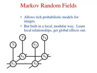
lcib.rutgers.edu Markov Random Field Basics • Goal: Inject knowledge that malignant glands are near malignant glands • Establish a graph connecting the glands • Let {a1, a2,…, aN}be the gland areas • Let {l1, l2,…, lN}be the gland labels with li{m,b}
lcib.rutgers.edu Markov Random Field Models • Prevalent parametric model (Ising) • Generic model used for its simplicity • Novel nonparametric model • Generated directly from image statistics
lcib.rutgers.edu Experiments • Experiment 1 Evaluate CAD gland classification performance • Experiment 2 Compare parametric (Ising) and nonparametric models • Dataset four H&E stained whole-mount histological sections at 10 micron
lcib.rutgers.edu Experiment 1: CAD Performance Area-based with MRF
lcib.rutgers.edu Experiment 2: Compare Ising and Nonparametric Model Gland Segmentation Nonparametric Gland Classification Ising Ising MRF Nonparametric MRF
lcib.rutgers.edu Concluding Remarks • First CAD system for detecting CaP in whole-mount histological images • Sensitivity of 0.8670 and specificity of 0.9524 • Requires 4-5 minutes on a 2100×3200 image using standard desktop PC • Introduced a novel nonparametric model for Markov Random Fields • Better performance than Ising model • Easily extended to other biological applications
lcib.rutgers.edu Acknowledgements • Wallace H. Coulter Foundation • New Jersey Commission on Cancer Research • National Cancer Institute • Society for Imaging and Informatics on Medicine • Life Science Commercialization Award
lcib.rutgers.edu The End
Gibbs Formulations • Generic Ising Model • Nonparametric Formulation
MRF Basics: Markov Properties • Let G={S,E}define a graph on N glands • Let y = {y1, y2,…, yN}be the gland areas • Let x = {x1, x2,…, xN}be the gland labels with xi{m,b} • Use maximum a posteriori estimation to obtain x • Simplify with Markov Property: p(xs|x-s)=p(xs|xr:rs) • Markov Property implies p(x) is a Gibbs distribution
Prostate Cancer • Among men in the US prostate cancer (Cap) is second most common cancer and the second leading cause of cancer-related death. • Histological analysis provides the definite test for CaP. • Analysis of whole mount histological sections (WMHSs) • Staging and grading of CaP • Ground truth for other modalities
Segmentation: Region Growing Internal Boundary (IB) Current Boundary (CB) Current Region (CR) 45 2 Iteration 132 126 178 14 3 4 5 1 boundary_measure = mean(IB)-mean(CB)
Quantitative Results • Review of algorithm • Segmentation • Classification using area (requires a probability threshold) • MRF Iteration • Initial conditions affect MRF results • ROC curve over varying thresholds Gland Classification Performance
Experiment 1: Gland Classification • Evaluate the ability to discriminate malignant from benign glands. • A gland whose centroid lies within the blue truth is considered cancerous, otherwise it is benign. • Training/test data consists of four slices using a leave-one-out training