Download
1 / 25
250 likes | 600 Views
Segmentation of Dermatological Images using Mixture Models and Markov Random Fields. Ji Tran Supervisor: Dr. David Squire. Overview. Introduction The Problem Definitions Aims Solution Results & Discussion Problems Encountered Conclusion Future Work References. Introduction.

E N D
Segmentation of Dermatological Images usingMixture Models and Markov Random Fields Ji Tran Supervisor: Dr. David Squire
Overview Introduction The Problem Definitions Aims Solution Results & Discussion Problems Encountered Conclusion Future Work References
Introduction Australia and many other countries in the world have very dry and sunny conditions. Many people each year die from skin cancer, and this statistic is rising steadily each year (6 percent). If skin cancer is detected early, the cure rate is high. Dermatologists have been trying different methods to aid in the detection of the rapid growing skin cancer threat.
The Problem Skin cancer cases are often misdiagnosed due to the lack of experience and knowledge of the different skin lesion types. Generic CBIRS exists, but their image processing techniques are not effective. Dermatologists are inconsistent and subjective in drawing borders around skin lesion due to the different levels of experience. Most image processing techniques employed are affected by noise, especially the presence of hair.
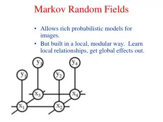
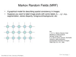
Definitions Content-Based Image Retrieval Systems (CBIRS) Image Segmentation Clustering Fuzzy Clustering Markov Random Field Gaussian Mixture Models
Aims Research and implement improved image segmentation models to produce better segmentation results for the dermatological images. Replace the existing skin lesion detection methods using image segmentation techniques. Implement segmentation algorithms that are resistant to noise (hair) as well as being independent of other pre-processing techniques. Study effective segmentation models that can be integrated into the foundation of a CBIRS tailored for the dermatological domain.
Solution • Implement the MRF segmentation model by (Deng & Clausi; 2004). • Extend the above algorithm to use a better distance criteria and a different MRF layout. • Compare the results of the two algorithms above against the classical clustering methods. • Because a skin lesion image consists of a skin lesion and background (healthy skin), we can segment the images using two clusters only using the RGB colour space (HSL has problems). • Build a visualisation tool to verify and validate the behaviours of the above segmentation models.
Classical Methods • Kmeans • Fuzzy Cmeans • Gaussian Mixture Models • Based on two common distance measures, Euclidean and Mahalanobis distance.
Classical Method Downfalls • They are easily affected by noise. • Clustering using the Euclidean distance (square distance) measure is not accurate as it does not consider the variances of the mixture models. • The methods that are based on the Mahalanobis distance do not model the region (spatial) properties of a data set. This leads to situations where the variances of a cluster shrinks to a thin band (GMM). Kmeans (E) Kmeans (M) Fuzzy Cmeans (E) Fuzzy Cmeans (M) GMM
MRF Segmentation Model – using a Variable Weighting Factor • Brief overview… • Consists of two main components. • A region component that captures the spatial relationships between pixels in their MRF. • A feature component that models the features in the feature data. • Uses a variable weighting factor to combine the two regions such that a good trade-off between the two is established.
MRF Segmentation Model – using a Variable Weighting Factor (cont.)
MRF Segmentation Model – using a Variable Weighting Factor (cont.)
MRF Segmentation Model – the Improved Model • We improved the previous method by using a full covariance to model the feature energy component of each data point. • The previous model used a simple 4 neighbourhood MRF. • Our model can use the 4 or 8 neighbourhood MRF, but a MRF size of 8 smooths the segmentation borders. • A β constant in front of the α(t) is used to control the smoothness.
Results & Discussion • General Images • The results generated by the classical methods used the default settings. While the MRF based segmentation models had the following settings: • K = 2, Final temp. = 0.1, Initial temp = 15, β = 0.1, c1 = 80, c2 = 1/K • Both the full covariance with an 8 neighbourhood MRF and the single variance with a 4 neighbourhood MRF models were used. • Dermatological Images • The same settings were used, but we have found that using the β = 0.01 works best for the dermatological images. • For the MRF based segmentation models, 25 EM iterations and 100 simulated annealing steps were used.
Results – General Images “…here’s Johnny!...” Kmeans (E) Kmeans (M) Fuzzy Cmeans (E) Fuzzy Cmeans (M) GMM MRF SV 4N MRF FC 8N Original
Results – Dermatological Images • Malignant Melanoma Kmeans (E) Kmeans (E) MRF SV 4N Fuzzy Cmeans (E) Fuzzy Cmeans (M) MRF FC 8N GMM
Results – Dermatological Images (cont.) • Dermal Naevus 1 Kmeans (E) Kmeans (M) MRF SV 4N Fuzzy Cmeans (E) Fuzzy Cmeans (M) MRF FC 8N GMM
Results – Dermatological Images (cont.) • Same dermatological images using the β value of 0.01. • By changing the β value, the influence of the region and the feature components will have different effects. • A small influence on the region side will pay more attention to the features, where a feature data being different to the Gaussian its label belongs to will be penalised with a high cost, but less noise is removed. Similarly, less influence on the feature component will generate smoother segments where the region component dominates, but finer details are lost. SV 4N MRF FC 8N MRF
MRF Segmentation Model – Effects of simulated annealing • The simulated annealing phase of the MRF based segmentation model is very important, especially in removing the presence of noise. • Even with the right temperature settings, if the simulated annealing phase cools too quickly (not enough SA Steps), the estimated values won’t get enough time to settle properly, which results in undesirable results. • An example is shown in the right with the standard MRF settings for the single variance version. 5 SA Steps 50 SA Steps 100 SA Steps
Convergence Rate of the segmentation models Note: The MRF based segmentation models both do 100 simulated annealing steps at each EM iteration
Problems Encountered Limited access to the online dermatological image database Mathematics exception handling Singular covariance matrix problems No access to the dermatologists Because of this, we cannot prove that our results are entirely correct.
Conclusion The new MRF segmentation algorithms are clearly much better than the classical methods. Noise removal is definitely a strong feature of the model. The downside of using the MRF based approach is that it is of the tens and hundreds times slower than the classical methods. For real time use such as in the medical clinic, this is not desirable. Although the MRF based approaches are slow, it does do a good job in detecting the skin lesion as well as removing the presence of hair structures. This is beneficial for the study of dermatological images.
Future Work Integrate the MRF based image segmentation algorithms into a CBIRS specifically for the dermatology domain. Improve the efficiency of the MRF based segmentation algorithm. Rewrite the segmentation algorithms to utilise the power of parellel computers/clusters. Introduce texture features into the new MRF segmentation model.
References • Deng, H. and Clausi, D. A. (2004). Unsupervised image segmentation using a simple MRF model with a new implementation scheme, Pattern Recognition 37: 2323–2335. • Jain, A. K., Murty, M. N. and Flynn, P. J. (1999). Data clustering: a review, ACM Computing Surveys (CSUR) 31: 1–60. • …and 55 more in the final thesis. MRF SV 4N