Download
1 / 16
160 likes | 286 Views
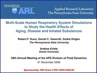
Multi-Scale Human Respiratory System Simulations to Study the Health Effects of Aging, Disease and Inhaled Substances Robert F. Kunz, Daniel C. Haworth, Gulkiz Do ğ an The Pennsylvania State University Andres Kriete Drexel University

E N D
Multi-Scale Human Respiratory System Simulations to Study the Health Effects of Aging, Disease and Inhaled Substances Robert F. Kunz, Daniel C. Haworth, Gulkiz Doğan The Pennsylvania State University Andres Kriete Drexel University 59th Annual Meeting of the APS Division of Fluid Dynamics 21 November 2006 Sponsorship: NIH Grant 5-R01-ES014483-02
Motivation • Goal 1: Develop, couple, apply, validate imaging and physics modeling of resolvable and sub-resolvable scales in human respiration • Goal 2: Provide clinical-time-scale (hours) diagnostic information for disease and injury assessment. • Step 1: Develop semi-automated end-to-end medical image through CFD analysis toolkit • Step 2: CFD modeling: • Unsteady • Multiphase/species • Full resolution for upper branches • Q1D for lower branches • Bulk modeling in respiratory units for volume, aerodynamic loss, deposition, gas exchange
Medical Imaging • CT scans of diseased patients • Clinical image standard: 3D sgi TIFF, DICOM header • Pixel dimension 0.6 x 0.6 mm, slice separation 0.8 mm. • Amira for image visualization and image processing • MEVISLab for DICOM headers for voxel sizes
Lobe Segmentation • Lobes needed for volume filling branching algorithm. • Fissures separating lung lobes are visible by a proper color map segment by following features/curves within 2-D transverse slices + follow anatomic features • Volume and surface of each lobe are calculated: • Smoothing, island removal, hole filling • STL export Right Left
Airway Tree Segmentation 81 year old male patient • Automated region growing based algorithm: • Threshold, colormap and interpolation features • Maximum generation to be segmented accurately depends on CT scan quality and the lung’s physical character for particular patient. • STL export
Thinning • Skeleton of the airways using distance map based thinning algorithm automated using an Amira script • Output node coordinates of skeleton, connectivity and local diameters along each segment. • @ one minute on a PC.
Partitioning • In-house code to automatically partition airway tree • Generation number of surface elements defined using ADT search to skeleton topology • Allows truncation of airway tree at desired location. • Enables automated assignment of model attributes (grid resolution, deposition efficiency) at different generations in grid generator and CFD code
Truncation • Same in-house code automatically truncates the airway trees and assigns the boundary conditions to truncated airways for fully-resolved 3D CFD calculations. After truncation at 0.85 through 5th generation Before truncation
Automated Octree Based Grid Generation • HARPOON for grid generation of both lobe and airway trees. Fully automated (batch mode) and quite fast: 1 million cells @ 1 minute on a PC. • Hexahedral dominant Cartesian meshes • “Hanging nodes” requires CFD code support for arbitrary polyhedra • Grid resolution is adapted by assigning different surface cell sizes to different generations • Prism layers as postprocessing step
Upper branch grid Cut plane xy Cut plane xz Automated Octree Based Grid Generation
Lower Branch Modeling • Another in-house code to reconstruct the tracheobronchial tree from each truncated airway down to respiratory units in each lobe. • A volume-filling algorithm has been devised such that the resulting spatial distribution of respiratory units is statistically uniform within each lobe.
Lower Branch Modeling • Branching algorithm reproduces geometric statistics (diameters, lengths, branching angles) of a human tracheobronchial tree. • Typical tracheobronchial trees represent 22-23 generations, contain 70,000 - 100,000 branches; @ 1/2 are terminal branches ending at a respiratory unit.
CT SCANS AMIRA UPPER AIRWAYS STL LOBE STL MASTER SCRIPT (today) UPPER AIRWAYS SKELETON HARPOON GEO_LUNG LOBE TETRA MESH MASTER SCRIPT (tomorrow) PARTITIONED AND TRUNCATED STL BRANCHING CODE HARPOON LOWER BRANCHES [input] [software] [in-house code][outputs] COBALT OCTREE-BASED-GRID FILE CFD Summary of Geometry Work
CFD Approach • Upper branches resolved @ O(105 - 106) elements • Lower branches modeled as Q1D pipe sections @ O(105) elements • Volume, mass flow rates conserved, friction factors modeled: • Flow losses in lower branches due to pipe wall shear modeled using “Q1D wall functions” • Flow losses in lower branches due to curvature/branching modeled using classical loss factors • Unstructured finite-volume CFD code – arbitrary polyhedral element support • Interphase coupling for Eulerian multiphase modeling (particle transport) • Parallelized for rapid execution on cluster computers (2-8 hours)
CFD Approach • Ensemble averaged n-field mass and momentum conservation laws in Cartesian tensor form: