Download
1 / 7
90 likes | 385 Views
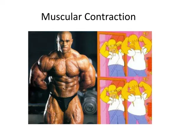
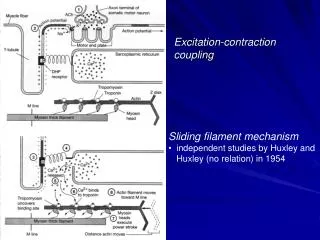
Excitation-contraction coupling. Sliding filament mechanism independent studies by Huxley and Huxley (no relation) in 1954. Excitation-Contraction. contraction occurs when Ca 2+ binds with troponin C each cycle takes ~50 msec and sarcomere shortens ~10 nm

E N D
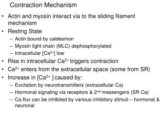
Excitation-contraction coupling • Sliding filament mechanism • independent studies by Huxley and Huxley (no relation) in 1954
Excitation-Contraction • contraction occurs when Ca2+ binds with troponin C • each cycle takes ~50 msec and sarcomere shortens ~10 nm • contraction ceases when Ca2+ is removed from sarcoplasm
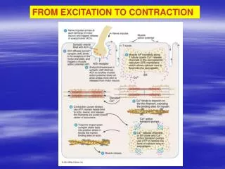
ATP Hydrolysis: Overview • in resting state, ATP is partially hydrolyzed • myosin head binds to actin filament when binding becomes available • ATP is completely hydrolyzed, and Pi and ADP are released from myosin head • actomyosin complex is broken when ATP binds to myosin
Maintenance of [ATP]i myosin ATPase ATP + H2O <—————> ADP + Pi + H+ creatine kinase PCr + ADP + H+ <—————> Cr + ATP adenylate kinase ADP + ADP <—————> ATP + AMP
ATP Hydrolysis: Weak Binding Phase • Transition 1 – A-M binding; ATP enters myosin ATP pocket • Transition 2 – ATP weakens A-M complex, myosin detaches • Transition 3 – ATP partially hydrolyzed • Transition 4 – myosin weakly reattaches to next actin molecule
ATP Hydrolysis: Strong Binding Phase • Transition 5 – Pi released from myosin ATP pocket; myosin cleft closes, strengthens binding • Transition 6 – power stroke of myosin head • Transition 7 – ADP released (rate-limiting step)
Transition 2 Transition 1 Transition 3 Weak Binding Transitions 6-7 Strong Binding Transition 5