Download
1 / 29
290 likes | 597 Views
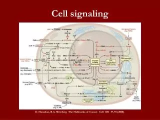
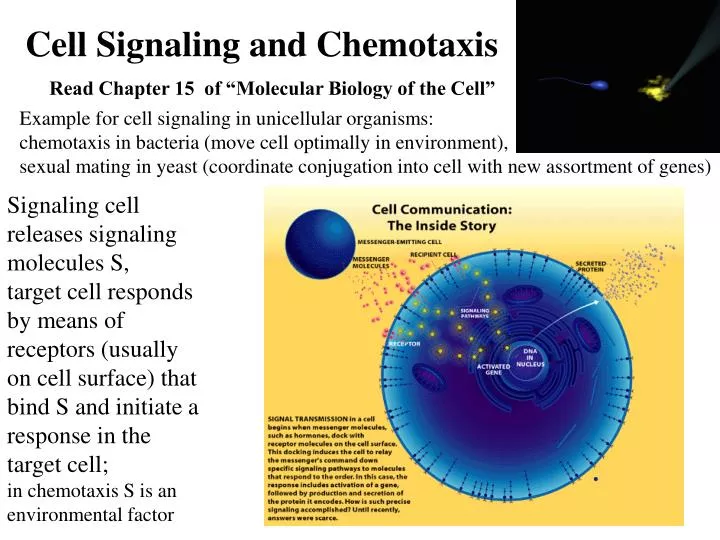
Cell Signaling and Chemotaxis. Read Chapter 15 of “Molecular Biology of the Cell”. Example for cell signaling in unicellular organisms: chemotaxis in bacteria (move cell optimally in environment), sexual mating in yeast (coordinate conjugation into cell with new assortment of genes).

E N D
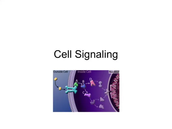
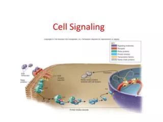
Cell Signaling and Chemotaxis Read Chapter 15 of “Molecular Biology of the Cell” Example for cell signaling in unicellular organisms: chemotaxis in bacteria (move cell optimally in environment), sexual mating in yeast (coordinate conjugation into cell with new assortment of genes) Signaling cell releases signaling molecules S, target cell responds by means of receptors (usually on cell surface) that bind S and initiate a response in the target cell; in chemotaxis S is an environmental factor
Sexual Mating in Yeast (coordinate conjugation into cell with new assortment of genes) When a haploid individual is ready to mate, it releases a peptide mating factor that signals cells of the opposite mating types to stop proliferating and prepare to conjugate; the subsequent fusion of two haploid cells of the opposite mating types produces two diploid cells, which then undergo meiosis and sporulate to generate haploid cells with a new assortment of genes (Alberts et al, Chpt. 15)
John D. Scott and Tony Pawson Scientific American before after Cell Signaling and Chemotaxis Read Chapter 15 of “Molecular Biology of the Cell” Example for cell signaling in unicellular organisms: chemotaxis in bacteria (move cell optimally in environment), sexual mating in yeast (coordinate conjugation into cell with new assortment of genes) Signaling cell releases signaling molecules S, target cell responds by means of receptors (usually on cell surface) that bind S and initiate a response in the target cell; in chemotaxis S is an environmental factor
G-proteins are Signal Transducers MissingG-protein signal signal Receptor Amplifier Receptor G-protein Amplifier Cell membrane Cytosol No Biological effect Biological effect Malfunctioning G-proteins disturb the intracellular signaling pathways, altering normal cell functions. G-proteins transmit and modulate signals in cells. They can activate different cellular amplifier systems. GTP GDP + Pi + 7.3 kcal/mol Ras switch I • smallest G-protein (189 residues, 21KDa mass) • acts as a molecular switch • cycles between an active (GTP-bound) and an inactive (GDP-bound) state • major conformational changes during the signaling cycle take place in the switch I and switch II regions • switching activity regulated by GAP and GEF proteins • activated forms of Ras genes are found in 30% of human tumors. switch II
Ras GDP GTP Ras GTP signal IN Signaling Cycle of Ras OFF R-state GTP hydrolysis is induced by GAP protein Pi GTP hydrolysis Guanine nucleotide Exchange Factor GTPase Activating Protein GEF Conformationalchange GAP GDP Exchange of GDP for GTP is catalyzed by GEF protein T-state ON signal OUT
GDP GTP Mechanical Cycle of Ras/Spring Ras Ras/GTP hydrolysis, induced by GAP, leads to Ras/GDP in T-state GTP hydrolysis GTP Ras/GDP Pi Ras/GDP evolves irreversibly and spontaneously fromT-statetoR-state GAP Ras Ras/GDP GDP T-state ~ 1ns Ras separates from GAP, then exchanges GDP for GTP, and the reverse R-to-T transition takes place GAP TRtransiton Ras GDP R-state
What Happens After GTP Hydrolysis? Switch I RAS/GDP RAS/GTP small fluctuations T-state strong fluctuations R-state Switch II Switch II helix “melts” altering the contact area of RAS!
The Role of Modules in Signaling John D. Scott and Tony Pawson Scientific American
Scaffolds Speed Signal Transmission John D. Scott and Tony Pawson Scientific American
Neutrophils are our body's first line of defense against bacterial infections. After leaving nearby blood vessels, these cells recognize chemicals produced by bacteria in a cut or scratch and migrate "toward the smell". The above neutrophils were placed in a gradient of fMLP (n formyl methionine- leucine- phenylalanine), a peptide chain produced by some bacteria. The cells charge out like a "posse" after the bad guys. http://www.cellsalive.com/chemotx.htm
Chemotaxis of neutrophil chasing a bacterium (http://www.hopkinsmedicine.org/cellbio/devreotes/movies.html) This video is taken from a 16mm movie made in the 1950s by the late David Rogers at Vanderbilt University. It was given to me via Dr. Viktor Najjar, Professor Emeritus at Tufts University Medical School and a former colleague of Rogers. It depicts a human polymorphonuclear leukocyte (neutrophil) on a blood film, crawling among red blood cells, notable for their dark color and principally spherical shape. The neutrophil is "chasing" Staphylococcus aureus microorganisms, added to the film. The chemoattractant derived from the microbe is unclear, but may be complement fragment C5a, generated by the interaction of antibodies in the blood serum with the complement cascade. Blood platelets adherent to the underlying glass are also visible. Notable is the characteristic asymmetric shape of the crawling neutrophils with an organelle-excluding leading lamella and a narrowing at the opposite end culminating in a "tail" that the cell appears to drag along. Contraction waves are visible along the surface of the moving cell as it moves forward in a gliding fashion. As the neutrophil relentlessly pursues the microbe it ignores the red cells and platelets. However, its leading edge is sufficiently stiff (elastic) to deform and displace the red cells it bumps into. The internal contents of the neutrophil also move, and granule motion is particularly dynamic near the leading edge. These granules only approach the cell surface membrane when the cell changes direction and redistributes its peripheral "gel." After the neutrophil has engulfed the bacterium, note that the cell's movements become somewhat more jerky, and that it begins to extend more spherical surface projections. These bleb-like protruberances resemble the blebs that form constitutively in the M2 melanoma cells missing the actin filament crosslinking protein filamin-1 (ABP-280) and may be telling us something about the mechanism of membrane protrusion. Written by Tom Stossel, June 22, 1999.
Genetics of Chemotactic Signaling System http://www.genome.ad.jp/kegg/pathway/eco/eco02030.html
Adaptation Model Adaptation in Chemotaxis slow slow intermediate fast
Summary of the experiments of Lumsden and Davies showing chemotaxis between neural tissue (trigeminal ganglion) and its target (whisker pad). The chemoattraction is specific for (A) the target and (B) the epithelial cells of the target. Moreover, the chemotactic ability of the whisker pad is specific for the trigeminal neurons.