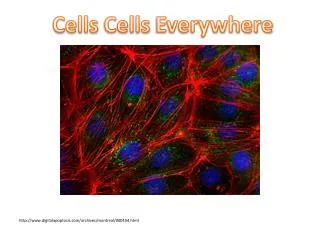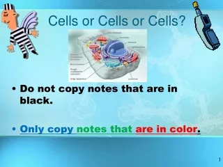
Cells
Cells . Cell Theory. Cells are the fundamental (smallest) units of life. All organisms are composed of cells. All cells come from preexisting cells. (Proved by Pasteur who disproved spontaneous generation in 1859.) Spontaneous Generation Formulated in 1838 by Schwann and Schleiden .

Cells
E N D
Presentation Transcript
Cell Theory • Cells are the fundamental (smallest) units of life. • All organisms are composed of cells. • All cells come from preexisting cells. (Proved by Pasteur who disproved spontaneous generation in 1859.) • Spontaneous Generation • Formulated in 1838 by Schwann and Schleiden.
Surface area to volume ratio • Cells have a large surface area to volume ratio. • The cell’s volume determines the rate of chemical activities over time. • The cell’s surface area determines the amount of substances a cell can take in an expel. • Because cells need to transport substances frequently, small size is essential. • How do you increase cell SA without increasing size?
SA • Increase the folding to increase SA. • The respiratory tract has the SA of a tennis court due to folding… • The intestines has the SA of a football field…. • The excretory system has large SA for N removal: • Ammonia in fish • Urea in humans and animals • Uric acid in birds and reptiles
Key Nutrient absorption Vein carrying blood to hepatic portal vessel Microvilli (brush border) Blood capillaries Villi and Microvilli on the interior of the small intestine Epithelial cells Muscle layers Epithelial cells Large circular folds Lacteal Villi Lymph vessel Villi Intestinal wall
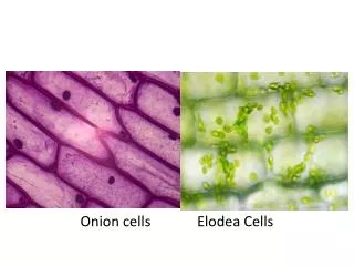
Since cells are small we need ocular assistance… Most cells are < 200 μm in size. Minimum resolution of human eye is 200 μm. Resolution is the distance apart that two objects must be in order for the eye to view them as distinct not a blur. Microscopes improve resolution.
Types of Microscopes • Light microscopes- glass lens and visible light to form a magnified image of an object. Magnifies about 1000x. • Electron microscopes-uses electromagnets to focus an electron beam. The beam is then directed to a fluorescent screen/photographic film to create a visible image. Magnifies about 1,000,000x. Can see subcellular.
Cell Similarities • 1. All cells have a cell membrane. (Phospholipidbilayer.) • 2. All cells contain DNA.
Cell Membrane Structure • 1. Phospholipids- are amphipathic. They line up to form a barrier from the water that is inside/outside the cell. • Remember Phosphate heads are (-) • Phosphates prevent hydration shells around each phospholipid. • 2. Proteins- are amphipathic. • A. Integral run through c.m. Function: structure and transport • B. Peripheral- on one side of c.m. Function: attachment of cytoskeleton and ECM
AmphipathicPhospholipids WATER Hydrophilic head Hydrophobic tail WATER
Hydrophilic region of protein AmphipathicProteins Phospholipid bilayer Hydrophobic region of protein
C.M. Proteins Cont. The proteins of the cell membrane can have several functions. • Molecule transport (Helps move food, water, or something across the membrane.) • Act as enzymes (To control metabolic processes.) • Cell to cell communication and recognition (So that cells can work together in tissues.) • Signal Receptors (To catch hormones or other molecules circulating in the blood.)
Signal Enzymes Membrane Protein Functions Receptor ATP Enzymatic activity Transport Signal transduction
Glyco- protein Membrane Protein Functions Attachment to the cytoskeleton and extra- cellular matrix (ECM) Cell-cell recognition Intercellular joining
C.M. Cont. • 3. Cholesterol- keeps c.m. flexibility. Also prevents plant cell membranes from freezing
Synthesis Question (U1,D12) • Question: All living cells have to have a cell membrane to remain living and intact. All cells are mostly water inside the cell. Cells live mainly in a watery environment. Water is a polar molecule. In four sentences or less, how does the presence of water on the inside and outside of a cell contribute to the structure of cell membranes? (5 Points) 1pt. Discussion of negative phosphorus atoms, of phospholipids being attracted to water and forming a barrier in the bi-layer. 1pt. Discussion of the bi-layer needed to prevent water from forming hydration shells • around the phospholipids. 1pt. Discussion of the fatty acid tails being protected sandwiched in-between the Phosphorus barriers. 1pt. Correct use of scientific terms. 1pt. Answer has no more than three sentences. (Following Directions.)
Prokaryotic Cells • “Kary” means kernel, in this case the nucleus. • Prokaryotes compose the Domains: Archaea and Bacteria. • Do not have membrane bound organelles. • Thought to have been the “first cells”
Prokaryotes • Can live in more diverse environments than eukaryotes. • Can sustain life on more diverse energy sources than eukaryotes. • Typically smaller than eukaryotes. • Tend to aggregate in chains or clusters.
Prokaryote Composition • 1. Plasma membrane regulating incoming/outgoing substances. • 2. Nucleiod- contains DNA, not a defined region • 3. Cytoplasm: (2 parts) • Cytosol-consists mostly of water with ions and water soluble molecules such as proteins • Insoluble particles including ribosomes. • Ribosomes are RNA and proteins.
Specialized Prokaryotic Features • Some prokaryotes developed specialized features. Why? • 1. Cell Walls- located exterior to the cell membrane. Dissimilar to plant cell walls • Cell walls typically contain peptidoglycan an amino sugar • Sometimes have an outer membrane. • Sometimes a capsule:= Slime layer made of polysaccharides; prevents drying out and aids in attachment to other cells (sickness)
Cell Walls, Internal Membranes, & Flagella and Pili • Plasma membrane will fold in to form specialized compartments for photosynthesis, cell division, or catabolic activities. • Structures of Movement • Flagella- made of protein flagellin, rotates like an axel for movement. • Pili- aka cilia, hairlike projections for movement
Cytoskeleton- helical structures just inside the plasma membrane. • Composed of proteins similar to actin. Actin makes up cytoskeleton in eukaryotes. • Cytoskeleton commonly found in rod shaped bacteria.
Let’s Practice Cell Review
Eukaryotic Cells • Eukaryotic cells ~10x bigger than prokaryotes. • Have membrane bound organelles. • Organelle can be membrane bound or not. • Organelles have a specific functions and shapes. • The role/function of an organelle is defined by the chemical reactions that take place
Cell fractionation and microscopy • Cell organelles first detected by light microscopes. • Cell fractionation-break down plasma membrane, organelles separate based on size/density. • Biochemical analysis can then be done to detect for certain macromolecules • Many organelles identical in plants and animals.
Nucleus • Typically the largest organelle in animal cells. • Site of DNA (chromatin vs. chromosome). • Nucleolus- site of RNA and ribosome synthesis.
Nuclear Structure • Surrounded by a double membrane called the nuclear envelope. • ~3500 nuclear pores exist in the envelope • The pores consist of over 100 different proteins • These proteins will aggregate in pore complexes of 8 proteins. • Small molecules can get into the pores, larger molecules need a nuclear localization signal. (chain of amino acids)
Figure 4.8 The Nucleus is Enclosed by a Double Membrane (Part 2)
Figure 4.8 The Nucleus is Enclosed by a Double Membrane (Part 1)
Ribosomes- NOT ORGANELLES • Function: protein synthesis per nucleic acid instructions • Location: • Attached to Endoplasmic reticulum-out of cell proteins, • free-floating in cytoplasm-in cell proteins • inside mitochondria and chloroplasts. • Composed of two different subunits: • rRNA- ribosomal RNA • Protein molecules (>50 different ones)
Endomembrane System • Made up of Endoplasmic Reticulum and Golgi Bodies/Apparatus • Vesicles serve as transporters of substances between the endomembrane structure and within the cell.