Download
1 / 14
480 likes | 1.87k Views
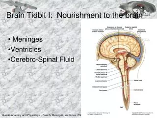
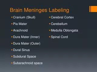
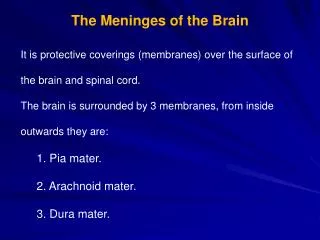
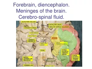
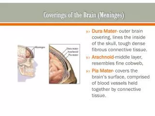
Coverings of the Brain (Meninges). Dura Mater- outer brain covering, lines the inside of the skull, tough dense fibrous connective tissue. Arachnoid -middle layer, resembles fine cobweb, Pia Mater- covers the brain’s surface, comprised of blood vessels held together by connective tissue.

E N D
Coverings of the Brain (Meninges) • Dura Mater- outer brain covering, lines the inside of the skull, tough dense fibrous connective tissue. • Arachnoid-middle layer, resembles fine cobweb, • Pia Mater- covers the brain’s surface, comprised of blood vessels held together by connective tissue.
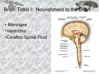
Subarachnoid Space • Between arachnoid and pia mater. Filled with cerebrospinal fluid- acts as a liquid shock absorber and source of nutrients for the brain.
Ventricles of the Brain • The brain contains four cavities filled with cerebrospinal fluid called cerebral ventricles • Right and left lateral ventricles • Third ventricle-behind and below the lateral ventricles • Fourth ventricle is below the 3rd in front of the cerebellum and behind the pons and medulla oblongota
Choroid Plexus • Network of blood vessels lining the ventricles which helps in the formation of cerebrospinal fluid.
Cerebrospinal Fluid • Forms inside ventricles of the brain • Serves as a liquid shock absorber protecting the brain and spinal cord. • Blood brain barrier- choroid plexus capillaries prevent substances (like drugs) from penetrating brain tissue this makes infections, like meningitis difficult to cure.
Lumbar Puncture • Removal of CSF from spinal canal, needle puncture between 3rd and 4th lumbar vertebrae.
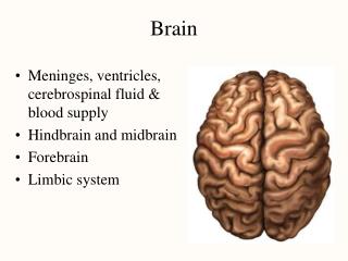
Cerebral Function- conscious thought, judgment, memory, reasoning, and will power Cerebrum • Largest part of the brain • Divided into R and L hemispheres by deep groove (longitudinal fissure) • Convolutions- elevated folds on the surface of the cerebrum, they increase the surface area of the brain • Sulci- fissure or grooves separating cerebral convolutions. Divided into four lobes- Frontal, Parietal, Occipital, and Temporal
Diencephalon • Located between cerebrum and midbrain • Composed of Thalamus and Hypothalamus • Vital functions of the hypothalamus: • Autonomic nervous control • Temperature control • Appetite control • Emotional state • Sleep control
Cerebellum • Located behind the pons and below the cerebrum • Composed of two hemispheres • Controls all body functions related to skeletal muscles, including: • Balance • Muscle tone • Coordination of muscle movements
Brain Stem • Made up of Pons, medulla and midbrain • Pathway for ascending and descending tracts • Pons – in front of cerebellum, between midbrain and medulla- contains center that controls respiration • Midbrain- vision and hearing • Medulla oblongota- bulb shaped structure between pons and spinal cord, inside the cranium above foramen magnum. Responsible for : • Heart rate • Blood pressure
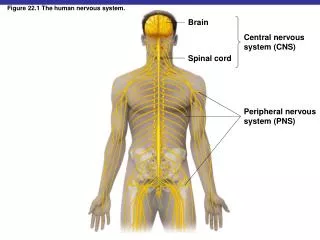
Spinal Cord • Begins at foramen magnum and continues down to 2nd lumbar vertebrae • White and soft, in spinal canal • Surrounded by cerebrospinal fluid • Functions as: • Reflex center • Conduction pathway to and from the brain
Limbic System • Part of the brain associated with emotional control, mood and memory • Includes the hypothalamus which is considered the brain of the brain.
Questions for 3.01 ppt BM two • Draw a picture of the brain and label all of the structures on the bubble map. • Color the bubble map and write the words in color. • Make sure your brain covers the entire page and each structure needs to be in color.