Brain and Cranial Nerves
530 likes | 1.2k Views
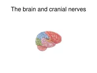
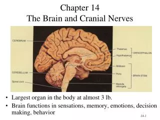
Brain and Cranial Nerves. Parts of the Brain. Parts of the Brain. CEREBRUM. DIENCEPHALON:. Thalamus. Hypothalamus. BRAIN STEM:. CEREBELLUM. Midbrain. Pons. Medulla oblongata. Spinal cord. (b) Sagittal section, medial view. Parts of the Brain. Meninges. Meninges. Ventricles.

Brain and Cranial Nerves
E N D
Presentation Transcript
Brain and Cranial Nerves
Parts of the Brain CEREBRUM DIENCEPHALON: Thalamus Hypothalamus BRAIN STEM: CEREBELLUM Midbrain Pons Medulla oblongata Spinal cord (b) Sagittal section, medial view
Cerebrum perception - sensory initiate voluntary movements memory learning understanding conscious behavior
Cerebrum • The lobes of the cerebrum correspond to the bones of the braincase which bear the same names. parietal frontal temporal occipital parietal frontal occipital temporal
Cerebrum Functional Areas 1. Sensory areas 2. Motor areas 3. Association area
Cerebrum Functional Areas Primary somatosensory area (1, 2, 3) Primary visual area (17) Primary auditory area (41, 42) Primary gustatory area (43) Primary olfactory area (28 - not visible) Primary motor area (4) Broca’s area (44, 45 left hemisphere) Somatosensory association area (5, 7) Prefrontal cortex (9, 10, 11, 12 -medial view only) Visual association area (18, 19) Auditory association Area (22) Wernicke’s area (22, 39, 40 - left hemisphere) Common integrative area (5, 7, 39, 40) Premotor area (6) Frontal eye field (8)
Cerebrum Motor/Sensory
Basal Nuclei • The basal nuclei also control subconscious contractions of skeletal muscles. Examples include automatic arm swings while walking and true laughter in response to a joke.
Limbic System • Encircling the upper part of the brain stem and the corpus callosum is a ring of structures on the inner border of the cerebrum and floor of the diencephalon that constitutes the limbic system. • The limbic system is sometimes called the “emotional brain” because it plays a primary role in promoting a range of emotions, including pleasure, pain, docility, affection, fear, and anger. • Together with parts of the cerebrum, the limbic system also functions in memory.
Brain Waves • Summing waves of different frequency produces some characteristic, and diagnostic patterns. • Alpha (10–12 Hz (cycles/sec) waves are present when awake but disappear during sleep. • Beta (14–30 Hz) waves are present with sensory input and mental activity when the nervous system is active. • Theta (4–7 Hz) waves indicate emotional stress or a brain disorder. • Delta (1–5 Hz) waves appear only during sleep in adults but indicate brain damage in an awake adult.
Brain Stem - Medulla Oblongata The medulla begins at the inferior border of the pons and extends to the foramen magnum. It contains all ascending and descending tracts extending between the spinal cord and cerebrum. The medulla contains nuclei which are regulators for vital body functions.
Medulla Oblongata Axons from the left pyramid cross over to the right and axons on the right cross over to the left (decussation of pyramids) – so that the left hemisphere of the brain controls the right side muscles, while the right hemisphere controls the left side.
Brain Stem -Pons • The pons lies directly above the medulla and anterior to the cerebellum (2.5 cm). It acts as a bridge connecting the spinal cord with the brain and parts of the brain with each other.
Brain Stem- Midbrain • The midbrain extends from the pons to the diencephalon. • The cerebral aqueduct passes through the midbrain connecting the 3rd ventricle above with the 4th ventricles below (both locations of CSF formation and circulation.)
Cerebellum Compares intention with actual performance Cerebrum initiates voluntary muscle contractions and notifies cerebellum. 2. Cerebellum gets information from proprioceptors. 3. Assesses information. 4. Dispatches “blueprint” for coordination to cerebrum
Diencephalon - Thalamus All sensory input Sort out information Major relay station for sensory ascending to sensory cortex, and inputs of subcortical motor nuclei and the cerebellum
Diencephalon - Hypothalamus 1. Autonomic control center 2. Center for emotional response 3. Body temperature regulation 4. Regulation of food intake 5. Regulation of water balance and thirst 6. Regulation of sleep-wake cycles 7. Control of endocrine system
Diencephalon- Epithalamus • Pineal gland • secretes melatonin during darkness • promotes sleepiness & sets biological clock • Habenular nuclei • emotional responses to odors
III. Oculomotor nerve IV. Trochlear nerve VI. Abducens nerve