Download
1 / 52
520 likes | 731 Views
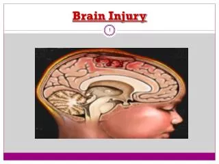
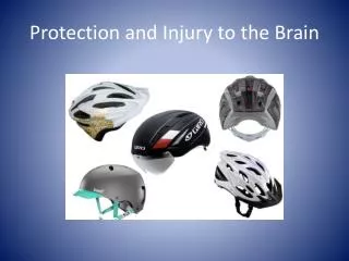
Protection and Injury to the Brain. Protection of the Brain. Nervous tissue is soft and easily injured. Several systems are in place to protect the brain. Meninges. The Meninges consist of three connective tissue membranes that cover the organs of the CNS. They have several functions:

E N D
Protection of the Brain Nervous tissue is soft and easily injured. Several systems are in place to protect the brain.
Meninges The Meninges consist of three connective tissue membranes that cover the organs of the CNS. They have several functions: • Cover and protect the CNS • Protect the blood vessels and venous sinuses • Contain cerebral spinal fluid • Form partitions within the skull
The three layers are • dura mater • arachnoid mater • pia mater (Think DAP)
Dura Mater The Dura Mater “tough mother” is composed of two layers of fibrous connective tissue. • The periosteal layer is in contact with the inner surface of the skull • The meningeal layer is the true external covering • They can form venous sinuses and dural septa
Dural septa and Dural venous sinuses. Superior sagittal sinus Skull Falx cerebri Scalp Straight sinus Occipital lobe Crista galli of the ethmoid bone Tentorium cerebelli Dura mater Falx Transverse sinus cerebelli Pituitary gland Cerebellum Temporal bone Arachnoid mater over medulla oblongata (a) Dural septa (b) Dural venous sinuses
Superior sagittal sinus Skull Falx cerebri Scalp Occipital lobe Tentorium cerebelli Dura mater Falx cerebelli Transverse sinus Cerebellum Temporal bone Arachnoid mater over medulla oblongata (b) Dural venous sinuses
Arachnoid Mater This is the middle layer and forms a loose brain covering (looks like a spiders web) It is separated from the dura mater by a potential space known as the subdural space.
Pia Mater “gentle mother” Has a rich supply of blood vessels. It is the only one which clings tightly to the brain and follows every convolution
Skull Epidural Space Dura Mater Subdural Space Arachnoid Subarachnoid space Pia Mater
Skin of scalp Periosteum Bone of skull Dura mater Periosteal Meningeal Superior sagittal sinus Arachnoid mater Pia mater Arachnoid villus Subdural space Blood vessel Falx cerebri (in longitudinal fissure only) Subarachnoid space
Conditions involving the Meninges • Meningitis is an inflammation of the meninges. • It can be, most commonly, of viralor bacterial origin • The bacterial form is potentially fatal while the viral form is self limited.
Signs and Symptoms • Most common signs are: • Headache • Nuchal rigidity (Can’t flex neck) • Altered mental status (acting strange)
Conditions involving the Meninges Brain bleeds can be of three types, epidural, subdural or subarachnoid.
They can wake up dead Epidural hemorrhage typically occurs from blood vessels bleeding into the space between the dura mater and the skull. • Often associated with head trauma • Characterized by arterial blood accumulating in the epidural space Individuals typically are lucid and then deteriorate rapidly due to bleeding from arteries.
They can wake up dead Subdural hemorrhage typically occurs from blood vessels bleeding into the space between the dura and the arachnoid layer. • Often associated with head trauma • Characterized by venous blood accumulating in the subdural space The onset of symptoms is gradual these include confusion and headache.
They can wake up dead Epidural Hematoma Subdural Hematoma
Conditions involving the Meninges Sub arachnoid hematoma occurs in the space between the arachnoid and pia matter. These typically present with stroke like symptoms. The most common sign is a “thunder clap” headache, vomiting and changes in the level of consciousness.
Conditions involving the Meninges Sub arachnoid hematoma usually occurs as a result of a ruptured blood vessel.
Cerebral Spinal Fluid (CSF) The CSF is found in on and around the brain and spinal column. It forms a cushion and allows the brain to float preventing it from crushing itself.
Cerebral Spinal Fluid (CSF) CSF has a makeup similar to blood plasma but has less protein.
Figure 12.26a Formation, location, and circulation of CSF. Superior sagittal sinus 4 Choroid plexus Arachnoid villus Interventricular foramen Subarachnoid space Arachnoid mater Meningeal dura mater Periosteal dura mater 1 Right lateral ventricle (deep to cut) Choroid plexus of fourth ventricle 3 Third ventricle 1 CSF is produced by the choroid plexus of each ventricle. Cerebral aqueduct Lateral aperture 2 CSF flows through the ventricles and into the subarachnoid space via the median and lateral apertures. Some CSF flows through the central canal of the spinal cord. Fourth ventricle Median aperture 2 Central canal of spinal cord 3 CSF flows through the subarachnoid space. (a) CSF circulation 4 CSF is absorbed into the dural venous sinuses via the arachnoid villi.
Cerebral Spinal Fluid The brain is made up of four ventricles. CSF is formed in these structures and flows through the CNS.
Cerebral Spinal Fluid • 1st/2nd= paired lateral ventricles (lie in cerebral hemispheres) [separated by septum pellucidum- transparent wall]
Cerebral Spinal Fluid • 1st/2nd= paired lateral ventricles (lie in cerebral hemispheres) [separated by septum pellucidum- transparent wall] • 3rd ventricle lies within diencephalon [connected to each lateral ventricles by interventricularforeamen]
Cerebral Spinal Fluid • 1st/2nd= paired lateral ventricles (lie in cerebral hemispheres) [separated by septum pellucidum- transparent wall] • 3rd ventricle lies within diencephalon [connected to each lateral ventricles by interventricularforeamen] • In midbrain is central cavity => cerebral aqueduct [connects 3rd/4th ventricle]
Cerebral Spinal Fluid • 1st/2nd= paired lateral ventricles (lie in cerebral hemispheres) [separated by septum pellucidum- transparent wall] • 3rd ventricle lies within diencephalon [connected to each lateral ventricles by interventricularforeamen] • In midbrain is central cavity => cerebral aqueduct [connects 3rd/4th ventricle] • 4th ventricle lies in the brain steam, dorsal to the pons
Cerebral Spinal Fluid CSF is formed in the choroid plexuses that hang is each ventricle.
Cerebral Spinal Fluid • A complication seen with the ventricle system is hydrocephalus. • This occurs when one of the aqueducts are blocked
Blood Brain Barrier This is a protective mechanism that helps to maintain a stable internal environment. This is to keep the neurons from firing uncontrollably when there is a slight shift in ion or water concentrations.
Blood Brain Barrier To reach the neurons, 3 layers must be passed. These are: • The endothelium of the capillary wall • The thick basal lamina surrounding each capillary • The “feet” or processes from the astrocytes touching the capillaries
Blood Brain Barrier The barrier is NOT effective against nonpolar compounds such as fats or gases, this why anesthetics, alcohol and nicotine can affect the brain.
Injuries • Major cause of death and disabilities world wide. • Major population are the young
Injury to the Brain Concussion which is a temporary alteration in brain function. This is typically caused by a blow to the head. Signs and symptoms usually include dizziness and mild headache.
Concussion • A study from McGill University in Montreal found 60 percent of college soccer players reported concussion symptoms at least once during a season. • The University of Pittsburgh’s Brain Trauma Research Center estimates 34 percent of college football players have had one concussion while 20 percent have endured multiple concussions.
Concussion • Neuro- Psychological testing is designed to test brain function and identify elements of cognitive damage and recovery that may not be discernible through self-reporting.
Stroke or CVA Cerebral vascular accidents or strokes are the single most common type of brain injury. This is brought about by a blockage of the arteries to the brain. Depending on where the blockage occurs, the CVA can go be mild or devastating.
This refers to a condition of sudden onset which is due to either a blocked artery or a broken (ruptured) artery. Stroke
Types of Strokes • Cerebral Infarction
Types of Strokes • Cerebral Infarction • Transient Ischemic Attack (TIA)
Types of Strokes • Cerebral Infarction • Transient Ischemic Attack (TIA) • Hemorrhagic
Cerebral Infarction • This is due to a blocked or partially blocked artery in the brain.
Transient Ischemic Attack • This is due to a blocked or partially blocked artery in the brain but the symptoms resolve on their own in 24 hours.
Hemorrhagic Stroke • This is due to a broken blood vessel.