Human Nervous System: Meninges, Diseases & Prevention
Explore the anatomy and diseases of the nervous system, including meningitis, encephalitis, and botulism. Learn about prevention methods and vaccination recommendations for various conditions.

Human Nervous System: Meninges, Diseases & Prevention
E N D
Presentation Transcript
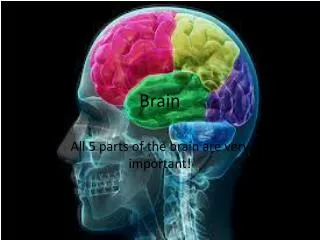
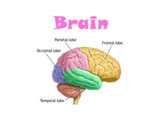
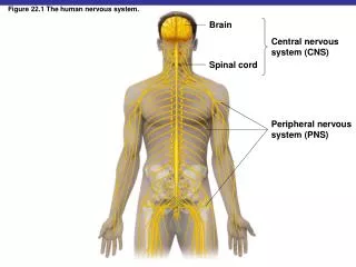
Figure 22.1 The human nervous system. Brain Central nervous system (CNS) Spinal cord Peripheral nervous system (PNS)
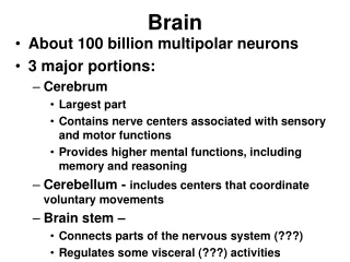
The Nervous System • Meninges protect brain and spinal cord • Dura mater: outermost layer • Arachnoid mater: middle layer • Subarachnoid space contains cerebrospinal fluid (CSF) • Pia mater: innermost layer • Blood–brain barrier
Figure 22.2 The meninges and cerebrospinal fluid. Skull bone Cerebrum Subarachnoid space (contains cerebrospinal fluid) Skull bone Dura mater Arachnoid mater Cranial meninges Pia mater Blood vessel Cerebrum Cerebellum Spinal cord Dura mater Spinalmeninges Arachnoid mater Pia mater Subarachnoid space of spinal cord Subarachnoid space (contains cerebrospinal fluid) Central canal
The Nervous System • Meningitis: inflammation of meninges • Encephalitis: inflammation of the brain • Meningoencephalitis: inflammation of both
Bacterial Meningitis • Initial symptoms of fever, headache, and stiff neck • Followed by nausea and vomiting • May progress to convulsions and coma • Diagnosis by Gram stain and latex agglutination of CSF
Figure 22.4 Spinal tap (lumbar puncture). Spinal needle is inserted, usually between the fourth and fifth lumbar vertebrae Cerebrospinal fluid Spinal cord Fourth lumbar vertebra L4 Cauda equina L5 Sample of cerebrospinal fluid Longitudinal section of the spine Fifth lumbar vertebra
Haemophilus influenzae Meningitis • Occurs mostly in children (6 months to 4 years) • Gram-negative aerobic bacteria, normal throat microbiota • Capsule antigen type b • Prevented by Hib vaccine
Neisseria Meningitis • Also called meningococcal meningitis • Caused by N. meningitidis • Gram-negative, aerobic cocci with a capsule • 10% of people are healthy nasopharyngeal carriers • Begins as throat infection, rash • Serotypes B, C, Y, W-135 • Serotype B & C in United States • Serotype A in Africa • Vaccination (A, C, Y, W-135 capsule) recommended for college students
Figure 22.3 Neisseria meningitis. N. meningitidis Cilia N. meningitidis
Streptococcus pneumoniae Meningitis • Also called pneumococcal meningitis • Caused by S. pneumoniae (a gram-positive diplococcus) • 70% of people are healthy nasopharyngeal carriers • Most common in children (1 month to 4 years) • Mortality: 30% in children, 80% in elderly • Prevented by vaccination
Listeriosis • Caused by Listeria monocytogenes • Gram-negative aerobic rod • Usually foodborne; it can be transmitted to fetus • Reproduce in phagocytes • Spread phagocyte-to-phagocyte
Figure 22.5 Cell-to-cell spread of Listeria monocytogenes, the cause of listeriosis. Listeria monocytogenes Macrophage Macrophage Pseudopod
Diseases in Focus: Meningitis and Encephalitis. Gram stain of cerebrospinal fluid.
Tetanus • Caused by Clostridium tetani • Gram-positive, endospore-forming, obligate anaerobe • Grows in deep wounds • Tetanospasmin released from dead cells blocks relaxation pathway in muscles • Prevention by vaccination with tetanus toxoid (DTaP) and booster (Td) • Treatment with tetanus immune globulin (TIG)
Botulism • Caused by Clostridium botulinum • Gram-positive, endospore-forming, obligate anaerobe • Intoxication comes from ingesting botulinal toxin • Botulinal toxin blocks release of neurotransmitter, causing flaccid paralysis • Prevention • Proper canning • Nitrites prevent endospore germination in sausages
Botulism • Treatment: supportive care and antitoxin • Infant botulism results from C. botulinum growing in intestines • Wound botulism results from growth of C. botulinum in wounds
Botulinal Types • Type A toxin • 60–70% fatality • Found in CA, WA, CO, OR, NM • Type B toxin • 25% fatality • Europe and eastern United States • Type E toxin • Found in marine and lake sediments • Pacific Northwest, Alaska, Great Lakes area
Leprosy • Also called Hansen’s disease • Caused by Mycobacterium leprae • Acid-fast rod that grows best at 30°C • Grows in peripheral nerves and skin cells • Transmission requires prolonged contact with an infected person
Leprosy • Tuberculoid (neural) form: loss of sensation in skin areas; positive lepromin test • Lepromatous (progressive) form: disfiguring nodules over body; negative lepromin test
Figure 22.9 Leprosy lesions. Tuberculoid (neural) leprosy Lepromatous (progressive) leprosy
Poliomyelitis (Polio) • Poliovirus • Transmitted by ingestion • Initial symptoms: sore throat and nausea • Viremia may occur; if persistent, virus can enter the CNS • Destruction of motor cells and paralysis occurs in <1% of cases • Prevention: vaccination (enhanced-inactivated polio vaccine)
Figure 22.11 Worldwide annual incidence of poliomyelitis. 7,000 6,000 2,000 5,000 1,000 4,000 Number of confirmed polio cases 0 2008 2010 2009 3,000 2,000 1,000 0 1999 2000 2001 2002 2003 2004 2005 2006 2007 2008 2009 2010 Year
Rabies • Caused by the rabies virus • Transmitted by animal bite • Furious rabies: animals are restless, then highly excitable • Paralytic rabies: animals seem unaware of surroundings
Figure 22.12 Pathology of rabies infection. Virus reaches brain and causes fatal encephalitis. Virus ascends spinal cord. Virus enters salivary glands and other organs of victim. Virus moves up peripheral nervous system to CNS. Virus replicates in muscle near bite and enters neuron of peripheral nervous system. Virus enters tissue from saliva of biting animal.
Figure 22.13 Reported cases of rabies in animals. Areas of the United States in which rabies predominates in certain wildlife species. Rabies-infected bats were reported in 47 of the 48 contiguous states. In eastern states in which raccoons are the predominant rabies-infected animal, many cases were also reported in foxes and skunks. KEY Skunk Raccoon Fox Fox and skunk
Figure 22.13 Reported cases of rabies in animals. Rabies cases in various wild and domestic animals in the United States. Rabies in domestic animals such as dogs and cats is uncommon because of high vaccination rates. Raccoons, skunks, and bats are the animals most likely to be infected with rabies. Most human cases are caused by bites of bats. Worldwide, most human cases are caused by bites of dogs. Raccoons 35.5% Bats 24.8% Skunks 24.5% Foxes 7.7% Cats 4.6% Dogs 1.2% KEY Cattle 1.1% Wild Horses/mules 0.6% Domestic 0 10 20 30 40 50
Clinical Focus: A Neurological Disease, Figure B. Silver-haired bat.
Rabies Virus • Virus multiplies in skeletal muscles and then brain cells, causing encephalitis • Initial symptoms may include muscle spasms of the mouth and pharynx and hydrophobia
Prevention of Rabies • Preexposure prophylaxis: injection of human diploid cells vaccine (HDCV) • Postexposure treatment: vaccine plus rabies immune globulin (RIG)
Cryptococcus neoformans Meningitis • Also called cryptococcosis • Soil fungus associated with pigeon and chicken droppings • Transmitted by the respiratory route; spreads through blood to the CNS • Mortality up to 30% • Treatment: amphotericin B and flucytosine
Chronic Fatigue Syndrome • Also called myalgic encephalomyelitis (ME) • Unexplained fatigue that lasts at least 6 months, plus four of these symptoms: • Sore throat • Tender lymph nodes • Muscle pain • Pain in multiple joints • Headaches • Unrefreshing sleep • Malaise after exercise • Impaired short-term memory or concentration
Chronic Fatigue Syndrome • Experimental treatment promotes antiviral interferons